• 文献检索
  • 文档翻译
  • 深度研究
  • 学术资讯
  • Suppr Zotero 插件Zotero 插件
  • 邀请有礼
  • 套餐&价格
  • 历史记录
应用&插件
Suppr Zotero 插件Zotero 插件浏览器插件Mac 客户端Windows 客户端微信小程序
定价
高级版会员购买积分包购买API积分包
服务
文献检索文档翻译深度研究API 文档MCP 服务
关于我们
关于 Suppr公司介绍联系我们用户协议隐私条款
关注我们

Suppr 超能文献

核心技术专利:CN118964589B侵权必究
粤ICP备2023148730 号-1Suppr @ 2026

文献检索

告别复杂PubMed语法,用中文像聊天一样搜索,搜遍4000万医学文献。AI智能推荐,让科研检索更轻松。

立即免费搜索

文件翻译

保留排版,准确专业,支持PDF/Word/PPT等文件格式,支持 12+语言互译。

免费翻译文档

深度研究

AI帮你快速写综述,25分钟生成高质量综述,智能提取关键信息,辅助科研写作。

立即免费体验

锥形束计算机断层扫描与多层螺旋计算机断层扫描中物体位置对密度测量的影响。

Effect of object location on the density measurement in cone-beam computed tomography versus multislice computed tomography.

作者信息

Eskandarloo Amir, Abdinian Mehrdad, Salemi Fatemeh, Hashemzadeh Zahra, Safaei Mehran

机构信息

Department of Oral and Maxillofacial Radiology, School of Dentistry, Hamedan University of Medical Science, Hamedan, Iran.

出版信息

Dent Res J (Isfahan). 2012 Dec;9(Suppl 1):S81-7.

PMID:23814567
原文链接:https://pmc.ncbi.nlm.nih.gov/articles/PMC3692205/
Abstract

BACKGROUND

Bone density measurement in a radiographic view is a valuable method for evaluating the density of bone quality before performing some dental procedures such as, dental implant placements. It seems that Cone-Beam Computed Tomography (CBCT) can be used as a diagnostic tool for evaluating the density of the bone, prior to any treatment, as the reported radiation dose in this method is minimal. The aim of this study is to investigate the effect of object location on the density measurement in CBCT versus Multislice computed tomography (CT).

MATERIALS AND METHODS

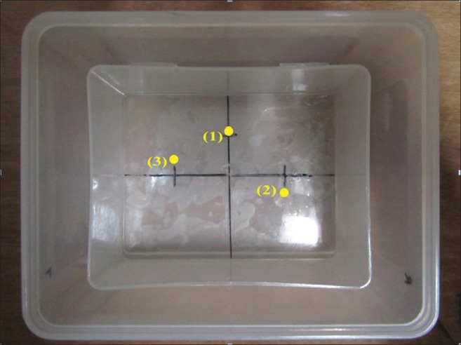
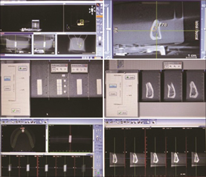
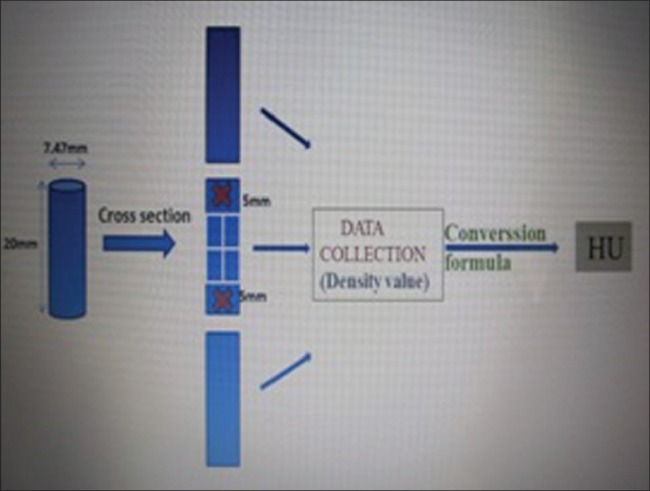
In an experimental study, three samples with similar dimensions, but different compositions, different densities (Polyethylene, Polyamide, Polyvinyl Chloride), and three bone pieces of different parts of the mandibular bone were imaged in three different positions by CBCT and Multislice CT sets. The average density value was computed for each sample in each position. Then the data obtained from each CBCT was converted to a Hounsfield unit and evaluated using a single variable T analysis. A P value <0.05 was considered to be significant.

RESULTS

The density in a Multislice CT is stable in the form of a Hounsfield Number, but this density is variable in the images acquired through CBCT, and the change in the position results in significant changes in the density. In this study, a statistically significant difference (P value = 0.000) has been observed for the position of the sample and its density in CBCT in comparison to Multislice CT.

CONCLUSIONS

Density values in CBCT are not real because they are affected by the position of the object in the machine.

摘要

背景

在进行某些牙科手术(如种植牙植入)之前,通过X线摄影视图测量骨密度是评估骨质密度的一种有价值的方法。锥形束计算机断层扫描(CBCT)似乎可以用作在任何治疗之前评估骨密度的诊断工具,因为该方法报告的辐射剂量极小。本研究的目的是调查物体位置对CBCT与多层计算机断层扫描(CT)中密度测量的影响。

材料与方法

在一项实验研究中,对三个尺寸相似但成分不同、密度不同(聚乙烯、聚酰胺、聚氯乙烯)的样本以及下颌骨不同部位的三块骨块,通过CBCT和多层CT设备在三个不同位置进行成像。计算每个样本在每个位置的平均密度值。然后将从每个CBCT获得的数据转换为亨氏单位,并使用单变量T分析进行评估。P值<0.05被认为具有显著性。

结果

多层CT中的密度以亨氏数的形式稳定,但通过CBCT获取的图像中的这种密度是可变的,并且位置的变化会导致密度的显著变化。在本研究中,与多层CT相比,观察到CBCT中样本位置及其密度存在统计学显著差异(P值=0.000)。

结论

CBCT中的密度值不真实,因为它们受物体在机器中的位置影响。

https://cdn.ncbi.nlm.nih.gov/pmc/blobs/8386/3692205/baaa9cfabd17/DRJ-9-81-g003.jpg
https://cdn.ncbi.nlm.nih.gov/pmc/blobs/8386/3692205/36e5aff3a1cb/DRJ-9-81-g001.jpg
https://cdn.ncbi.nlm.nih.gov/pmc/blobs/8386/3692205/04523d8821df/DRJ-9-81-g002.jpg
https://cdn.ncbi.nlm.nih.gov/pmc/blobs/8386/3692205/baaa9cfabd17/DRJ-9-81-g003.jpg
https://cdn.ncbi.nlm.nih.gov/pmc/blobs/8386/3692205/36e5aff3a1cb/DRJ-9-81-g001.jpg
https://cdn.ncbi.nlm.nih.gov/pmc/blobs/8386/3692205/04523d8821df/DRJ-9-81-g002.jpg
https://cdn.ncbi.nlm.nih.gov/pmc/blobs/8386/3692205/baaa9cfabd17/DRJ-9-81-g003.jpg

相似文献

1
Effect of object location on the density measurement in cone-beam computed tomography versus multislice computed tomography.锥形束计算机断层扫描与多层螺旋计算机断层扫描中物体位置对密度测量的影响。
Dent Res J (Isfahan). 2012 Dec;9(Suppl 1):S81-7.
2
Bone quality evaluation at dental implant site using multislice CT, micro-CT, and cone beam CT.使用多层螺旋CT、显微CT和锥形束CT对牙种植部位进行骨质量评估。
Clin Oral Implants Res. 2015;26(1):e1-7. doi: 10.1111/clr.12315. Epub 2013 Dec 11.
3
Comparison of gray values of cone-beam computed tomography with hounsfield units of multislice computed tomography: An study.锥形束计算机断层扫描的灰度值与多层螺旋计算机断层扫描的亨氏单位比较:一项研究。
Indian J Dent Res. 2017 Jan-Feb;28(1):66-70. doi: 10.4103/ijdr.IJDR_415_16.
4
Accuracy and reliability of different cone beam computed tomography (CBCT) devices for structural analysis of alveolar bone in comparison with multislice CT and micro-CT.与多层螺旋CT和微型CT相比,不同锥形束计算机断层扫描(CBCT)设备用于牙槽骨结构分析的准确性和可靠性。
Eur J Oral Implantol. 2017;10(1):95-105.
5
Morphometric analysis of mandibular trabecular bone using cone beam computed tomography: an in vitro study.使用锥形束计算机断层扫描对下颌骨小梁骨进行形态计量学分析:一项体外研究。
Int J Oral Maxillofac Implants. 2010 Nov-Dec;25(6):1093-8.
6
How accurate is CBCT in measuring bone density? A comparative CBCT-CT in vitro study.锥形束计算机断层扫描(CBCT)在测量骨密度方面的准确性如何?一项CBCT与计算机断层扫描(CT)的体外对比研究。
Clin Implant Dent Relat Res. 2014 Aug;16(4):471-8. doi: 10.1111/cid.12027. Epub 2013 Jan 7.
7
Conventional multi-slice computed tomography (CT) and cone-beam CT (CBCT) for computer-assisted implant placement. Part I: relationship of radiographic gray density and implant stability.用于计算机辅助种植体植入的传统多层螺旋计算机断层扫描(CT)和锥束CT(CBCT)。第一部分:影像学灰度密度与种植体稳定性的关系。
Clin Implant Dent Relat Res. 2013 Dec;15(6):893-906. doi: 10.1111/j.1708-8208.2011.00436.x. Epub 2012 Jan 17.
8
Influence of cone beam CT scanning parameters on grey value measurements at an implant site.锥形束 CT 扫描参数对种植体部位灰度值测量的影响。
Dentomaxillofac Radiol. 2013;42(3):79884780. doi: 10.1259/dmfr/79884780. Epub 2012 Aug 29.
9
Bone density: comparative evaluation of Hounsfield units in multislice and cone-beam computed tomography.骨密度:多层螺旋 CT 和锥形束 CT 中 Hounsfield 单位的比较评估。
Braz Oral Res. 2012 Nov-Dec;26(6):550-6. doi: 10.1590/s1806-83242012000600011.
10
Evaluation of voxel values in mandibular cancellous bone: relationship between cone-beam computed tomography and multislice helical computed tomography.下颌骨松质骨体素值评估:锥形束计算机断层扫描与多层螺旋计算机断层扫描之间的关系
Clin Oral Implants Res. 2009 May;20(5):503-6. doi: 10.1111/j.1600-0501.2008.01672.x. Epub 2009 Feb 25.

引用本文的文献

1
Comparative analysis of bone density measurements by using multislice spiral and cone-beam computed tomography.多层螺旋CT与锥形束CT骨密度测量的对比分析
J Dent Sci. 2020 Sep;15(3):388-389. doi: 10.1016/j.jds.2019.09.008. Epub 2019 Dec 31.
2
Effect of field-of-view size on gray values derived from cone-beam computed tomography compared with the Hounsfield unit values from multidetector computed tomography scans.与多探测器计算机断层扫描的亨氏单位值相比,视野大小对锥形束计算机断层扫描得出的灰度值的影响。
Imaging Sci Dent. 2018 Mar;48(1):31-39. doi: 10.5624/isd.2018.48.1.31. Epub 2018 Mar 19.
3
The relationship between the bone characters obtained by CBCT and primary stability of the implants.

本文引用的文献

1
Comparison of Conventional and Standardized Bone Densitometry around Implants in Periapical Radiographs during a Three Months Period.根尖片上种植体周围常规骨密度测定与标准化骨密度测定在三个月期间的比较
Dent Res J (Isfahan). 2011 Winter;8(1):33-8.
2
Effects of orthodontic tooth movement on alveolar bone density.正畸牙齿移动对牙槽骨密度的影响。
Clin Oral Investig. 2012 Jun;16(3):679-88. doi: 10.1007/s00784-011-0552-9. Epub 2011 Apr 26.
3
Analysis of intensity variability in multislice and cone beam computed tomography.多层和锥形束 CT 强度变化分析。
CBCT获得的骨特征与种植体初期稳定性之间的关系。
Int J Implant Dent. 2015 Dec;1(1):3. doi: 10.1186/s40729-014-0003-x. Epub 2015 Feb 12.
4
Effect of low-level laser therapy after rapid maxillary expansion: a clinical investigation.快速上颌扩弓后低强度激光治疗的效果:一项临床研究。
Lasers Med Sci. 2016 Aug;31(6):1185-94. doi: 10.1007/s10103-016-1970-3. Epub 2016 May 28.
5
Change in Image Quality According to the 3D Locations of a CBCT Phantom.根据锥形束计算机断层扫描(CBCT)体模的三维位置变化的图像质量
PLoS One. 2016 Apr 19;11(4):e0153884. doi: 10.1371/journal.pone.0153884. eCollection 2016.
6
CBCT-based bone quality assessment: are Hounsfield units applicable?基于锥形束计算机断层扫描的骨质量评估:亨氏单位是否适用?
Dentomaxillofac Radiol. 2015;44(1):20140238. doi: 10.1259/dmfr.20140238.
Clin Oral Implants Res. 2011 Aug;22(8):873-9. doi: 10.1111/j.1600-0501.2010.02076.x. Epub 2011 Jan 18.
4
Prospective study to estimate mandibular cancellous bone density using large-volume cone-beam computed tomography.使用大容量锥形束计算机断层扫描对下颌松质骨密度进行前瞻性研究。
Clin Oral Implants Res. 2010 Dec;21(12):1309-13. doi: 10.1111/j.1600-0501.2010.01950.x.
5
Reliability of voxel values from cone-beam computed tomography for dental use in evaluating bone mineral density.体素值在锥形束 CT 牙科应用中评估骨密度的可靠性。
Clin Oral Implants Res. 2010 May;21(5):558-62. doi: 10.1111/j.1600-0501.2009.01896.x.
6
[Evaluation of the implant sites of palatal implants using cone beam computed tomography].[使用锥形束计算机断层扫描评估腭部种植体的植入部位]
Zhonghua Kou Qiang Yi Xue Za Zhi. 2009 Jul;44(7):404-8.
7
Bone quality assessment based on cone beam computed tomography imaging.基于锥束计算机断层扫描成像的骨质量评估。
Clin Oral Implants Res. 2009 Aug;20(8):767-71. doi: 10.1111/j.1600-0501.2008.01677.x. Epub 2009 May 27.
8
Effect of object location on the density measurement and Hounsfield conversion in a NewTom 3G cone beam computed tomography unit.物体位置对NewTom 3G锥形束计算机断层扫描设备中密度测量及亨氏单位转换的影响
Dentomaxillofac Radiol. 2008 Sep;37(6):305-8. doi: 10.1259/dmfr/65993482.
9
Implant planning and placement using optical scanning and cone beam CT technology.使用光学扫描和锥形束CT技术进行种植体规划与植入
J Prosthodont. 2008 Aug;17(6):476-81. doi: 10.1111/j.1532-849X.2008.00322.x. Epub 2008 May 9.
10
Density conversion factor determined using a cone-beam computed tomography unit NewTom QR-DVT 9000.使用锥形束计算机断层扫描设备NewTom QR-DVT 9000确定的密度转换因子。
Dentomaxillofac Radiol. 2006 Nov;35(6):407-9. doi: 10.1259/dmfr/55276404.