Department of Clinical Radiology, University Hospital Muenster, Muenster, Germany.
Br J Cancer. 2013 Aug 6;109(3):658-66. doi: 10.1038/bjc.2013.356. Epub 2013 Jul 9.
Novel treatment strategies in Ewing sarcoma include targeted cellular therapies. Preclinical in vivo models are needed that reflect their activity against systemic (micro)metastatic disease.
Whole-body magnetic resonance imaging (WB-MRI) was used to monitor the engraftment and dissemination of human Ewing sarcoma xenografts in mice. In this model, we evaluated the therapeutic efficacy of T cells redirected against the Ewing sarcoma-associated antigen GD2 by chimeric receptor engineering.
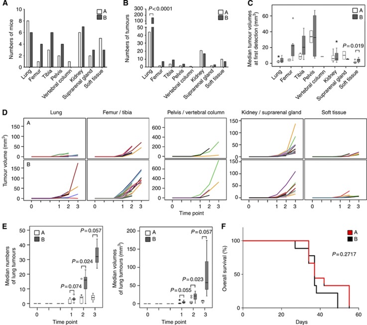
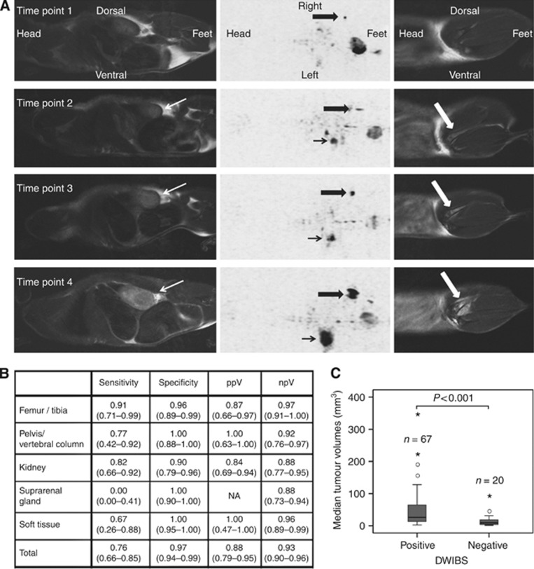
Of 18 mice receiving intravenous injections of VH-64 Ewing sarcoma cells, all developed disseminated tumour growth detectable by WB-MRI. All mice had lung tumours, and the majority had additional manifestations in the bone, soft tissues, and/or kidney. Sequential scans revealed in vivo growth of tumours. Diffusion-weighted whole-body imaging with background signal suppression effectively visualised Ewing sarcoma growth in extrapulmonary sites. Animals receiving GD2-targeted T-cell therapy had lower numbers of pulmonary tumours than controls, and the median volume of soft tissue tumours at first detection was lower, with a tumour growth delay over time.
Magnetic resonance imaging reliably visualises disseminated Ewing sarcoma growth in mice. GD2-retargeted T cells can noticeably delay tumour growth and reduce pulmonary Ewing sarcoma manifestations in this aggressive disease model.
尤文肉瘤的新型治疗策略包括靶向细胞疗法。需要能够反映其针对系统性(微)转移性疾病活性的临床前体内模型。
全身磁共振成像(WB-MRI)用于监测人尤文肉瘤异种移植物在小鼠中的植入和扩散。在该模型中,我们通过嵌合受体工程评估了针对尤文肉瘤相关抗原 GD2 的 T 细胞的治疗效果。
18 只接受静脉注射 VH-64 尤文肉瘤细胞的小鼠均发生了可通过 WB-MRI 检测到的弥散性肿瘤生长。所有小鼠均有肺部肿瘤,大多数小鼠还有骨、软组织和/或肾脏的其他表现。连续扫描显示体内肿瘤生长。背景信号抑制的全身扩散加权成像有效地可视化了肺外部位尤文肉瘤的生长。接受 GD2 靶向 T 细胞治疗的动物比对照组的肺部肿瘤数量更少,首次检测到的软组织肿瘤体积更低,并且随着时间的推移肿瘤生长延迟。
磁共振成像可可靠地可视化小鼠中播散性尤文肉瘤的生长。GD2 重定向 T 细胞可明显延迟肿瘤生长,并减少该侵袭性疾病模型中的肺部尤文肉瘤表现。