• 文献检索
  • 文档翻译
  • 深度研究
  • 学术资讯
  • Suppr Zotero 插件Zotero 插件
  • 邀请有礼
  • 套餐&价格
  • 历史记录
应用&插件
Suppr Zotero 插件Zotero 插件浏览器插件Mac 客户端Windows 客户端微信小程序
定价
高级版会员购买积分包购买API积分包
服务
文献检索文档翻译深度研究API 文档MCP 服务
关于我们
关于 Suppr公司介绍联系我们用户协议隐私条款
关注我们

Suppr 超能文献

核心技术专利:CN118964589B侵权必究
粤ICP备2023148730 号-1Suppr @ 2026

文献检索

告别复杂PubMed语法,用中文像聊天一样搜索,搜遍4000万医学文献。AI智能推荐,让科研检索更轻松。

立即免费搜索

文件翻译

保留排版,准确专业,支持PDF/Word/PPT等文件格式,支持 12+语言互译。

免费翻译文档

深度研究

AI帮你快速写综述,25分钟生成高质量综述,智能提取关键信息,辅助科研写作。

立即免费体验

使用边界分类法对黄斑OCT图像进行视网膜层分割

Retinal layer segmentation of macular OCT images using boundary classification.

作者信息

Lang Andrew, Carass Aaron, Hauser Matthew, Sotirchos Elias S, Calabresi Peter A, Ying Howard S, Prince Jerry L

机构信息

Department of Electrical and Computer Engineering, The Johns Hopkins University, Baltimore, MD 21218, USA.

出版信息

Biomed Opt Express. 2013 Jun 14;4(7):1133-52. doi: 10.1364/BOE.4.001133. Print 2013 Jul 1.

DOI:10.1364/BOE.4.001133
PMID:23847738
原文链接:https://pmc.ncbi.nlm.nih.gov/articles/PMC3704094/
Abstract

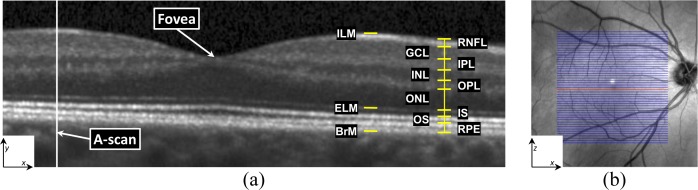
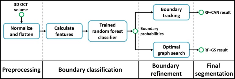
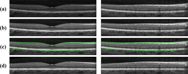
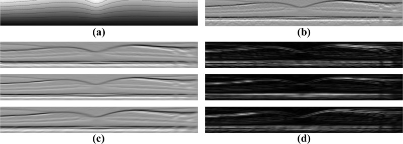
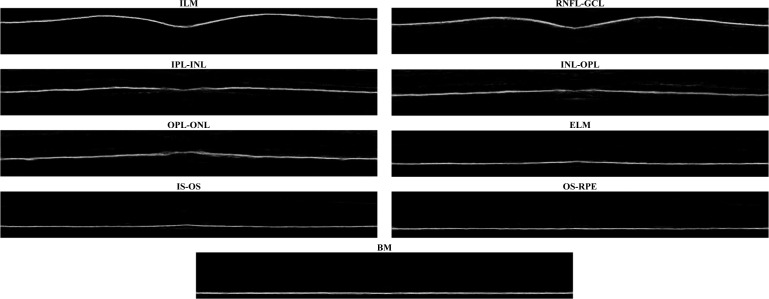
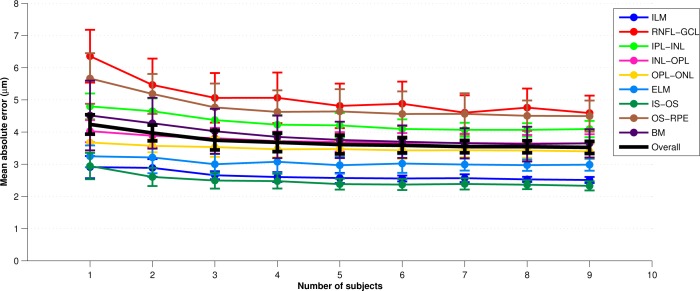
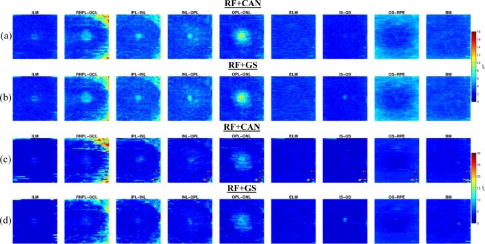
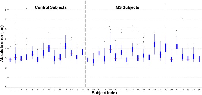
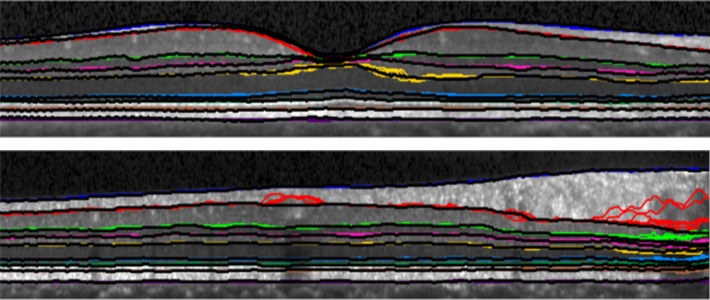
Optical coherence tomography (OCT) has proven to be an essential imaging modality for ophthalmology and is proving to be very important in neurology. OCT enables high resolution imaging of the retina, both at the optic nerve head and the macula. Macular retinal layer thicknesses provide useful diagnostic information and have been shown to correlate well with measures of disease severity in several diseases. Since manual segmentation of these layers is time consuming and prone to bias, automatic segmentation methods are critical for full utilization of this technology. In this work, we build a random forest classifier to segment eight retinal layers in macular cube images acquired by OCT. The random forest classifier learns the boundary pixels between layers, producing an accurate probability map for each boundary, which is then processed to finalize the boundaries. Using this algorithm, we can accurately segment the entire retina contained in the macular cube to an accuracy of at least 4.3 microns for any of the nine boundaries. Experiments were carried out on both healthy and multiple sclerosis subjects, with no difference in the accuracy of our algorithm found between the groups.

摘要

光学相干断层扫描(OCT)已被证明是眼科必不可少的成像方式,并且在神经学领域也显示出非常重要的作用。OCT能够对视神经乳头和黄斑处的视网膜进行高分辨率成像。黄斑视网膜层厚度提供了有用的诊断信息,并且已被证明与多种疾病的疾病严重程度指标密切相关。由于手动分割这些层既耗时又容易产生偏差,因此自动分割方法对于充分利用这项技术至关重要。在这项工作中,我们构建了一个随机森林分类器,用于分割通过OCT获取的黄斑立方图像中的八个视网膜层。随机森林分类器学习各层之间的边界像素,为每个边界生成准确的概率图,然后对其进行处理以确定边界。使用该算法,我们可以将黄斑立方体内包含的整个视网膜准确分割,对于九个边界中的任何一个,分割精度至少为4.3微米。我们对健康受试者和多发性硬化症受试者都进行了实验,发现两组之间我们算法的精度没有差异。

https://cdn.ncbi.nlm.nih.gov/pmc/blobs/3add/3704094/3256a62b75e2/boe-4-7-1133-g010.jpg
https://cdn.ncbi.nlm.nih.gov/pmc/blobs/3add/3704094/d72031dd6ba1/boe-4-7-1133-g001.jpg
https://cdn.ncbi.nlm.nih.gov/pmc/blobs/3add/3704094/1de1bfb7cbee/boe-4-7-1133-g002.jpg
https://cdn.ncbi.nlm.nih.gov/pmc/blobs/3add/3704094/2b7d261df19a/boe-4-7-1133-g003.jpg
https://cdn.ncbi.nlm.nih.gov/pmc/blobs/3add/3704094/2b9a431b6f05/boe-4-7-1133-g004.jpg
https://cdn.ncbi.nlm.nih.gov/pmc/blobs/3add/3704094/805a60838289/boe-4-7-1133-g005.jpg
https://cdn.ncbi.nlm.nih.gov/pmc/blobs/3add/3704094/ec2478c14253/boe-4-7-1133-g006.jpg
https://cdn.ncbi.nlm.nih.gov/pmc/blobs/3add/3704094/c2e14569a5b3/boe-4-7-1133-g007.jpg
https://cdn.ncbi.nlm.nih.gov/pmc/blobs/3add/3704094/ca727755d60d/boe-4-7-1133-g008.jpg
https://cdn.ncbi.nlm.nih.gov/pmc/blobs/3add/3704094/2290a7231cae/boe-4-7-1133-g009.jpg
https://cdn.ncbi.nlm.nih.gov/pmc/blobs/3add/3704094/3256a62b75e2/boe-4-7-1133-g010.jpg

相似文献

1
Retinal layer segmentation of macular OCT images using boundary classification.使用边界分类法对黄斑OCT图像进行视网膜层分割
Biomed Opt Express. 2013 Jun 14;4(7):1133-52. doi: 10.1364/BOE.4.001133. Print 2013 Jul 1.
2
Segmentation of retinal OCT images using a random forest classifier.使用随机森林分类器对视网膜光学相干断层扫描(OCT)图像进行分割。
Proc SPIE Int Soc Opt Eng. 2013 Mar 13;8669. doi: 10.1117/12.2006649.
3
Comparison of point estimates and average thicknesses of retinal layers measured using manual optical coherence tomography segmentation for quantification of retinal neurodegeneration in multiple sclerosis.使用手动光学相干断层扫描分割测量多发性硬化症中视网膜神经退行性变的视网膜层的点估计值和平均厚度的比较。
Curr Eye Res. 2013 Jan;38(1):224-8. doi: 10.3109/02713683.2012.722243. Epub 2012 Sep 6.
4
Layer boundary evolution method for macular OCT layer segmentation.用于黄斑光学相干断层扫描(OCT)层分割的层边界演化方法
Biomed Opt Express. 2019 Feb 4;10(3):1064-1080. doi: 10.1364/BOE.10.001064. eCollection 2019 Mar 1.
5
Automatic segmentation of OCT retinal boundaries using recurrent neural networks and graph search.使用递归神经网络和图搜索自动分割光学相干断层扫描(OCT)视网膜边界
Biomed Opt Express. 2018 Oct 26;9(11):5759-5777. doi: 10.1364/BOE.9.005759. eCollection 2018 Nov 1.
6
Multiple-object geometric deformable model for segmentation of macular OCT.用于黄斑光学相干断层扫描分割的多目标几何可变形模型
Biomed Opt Express. 2014 Mar 4;5(4):1062-74. doi: 10.1364/BOE.5.001062. eCollection 2014 Apr 1.
7
A supervised joint multi-layer segmentation framework for retinal optical coherence tomography images using conditional random field.基于条件随机场的视网膜光学相干断层扫描图像的监督式联合多层分割框架。
Comput Methods Programs Biomed. 2018 Oct;165:235-250. doi: 10.1016/j.cmpb.2018.09.004. Epub 2018 Sep 5.
8
Multi-phase level set algorithm based on fully convolutional networks (FCN-MLS) for retinal layer segmentation in SD-OCT images with central serous chorioretinopathy (CSC).基于全卷积网络的多阶段水平集算法(FCN-MLS)用于中心性浆液性脉络膜视网膜病变(CSC)的SD-OCT图像中的视网膜层分割。
Biomed Opt Express. 2019 Jul 15;10(8):3987-4002. doi: 10.1364/BOE.10.003987. eCollection 2019 Aug 1.
9
Normative Data and Minimally Detectable Change for Inner Retinal Layer Thicknesses Using a Semi-automated OCT Image Segmentation Pipeline.使用半自动光学相干断层扫描(OCT)图像分割流程获取的视网膜内层厚度的标准数据和最小可检测变化
Front Neurol. 2019 Nov 25;10:1117. doi: 10.3389/fneur.2019.01117. eCollection 2019.
10
Three-dimensional retinal imaging with high-speed ultrahigh-resolution optical coherence tomography.采用高速超高分辨率光学相干断层扫描技术的三维视网膜成像
Ophthalmology. 2005 Oct;112(10):1734-46. doi: 10.1016/j.ophtha.2005.05.023.

引用本文的文献

1
Combined optical coherence tomography and electroretinography system for imaging neurovascular coupling in the human retina.用于成像人类视网膜神经血管耦合的光学相干断层扫描与视网膜电图联合系统。
Neurophotonics. 2025 Jul;12(3):035004. doi: 10.1117/1.NPh.12.3.035004. Epub 2025 Aug 9.
2
An Artificial Intelligence Method for Phenotyping of OCT-Derived Thickness Maps Using Unsupervised and Self-supervised Deep Learning.一种使用无监督和自监督深度学习对光学相干断层扫描衍生的厚度图进行表型分析的人工智能方法。
J Imaging Inform Med. 2025 May 20. doi: 10.1007/s10278-025-01539-x.
3
Artificial intelligence in stroke risk assessment and management via retinal imaging.

本文引用的文献

1
Segmentation of retinal OCT images using a random forest classifier.使用随机森林分类器对视网膜光学相干断层扫描(OCT)图像进行分割。
Proc SPIE Int Soc Opt Eng. 2013 Mar 13;8669. doi: 10.1117/12.2006649.
2
Decision forests for tissue-specific segmentation of high-grade gliomas in multi-channel MR.用于多通道磁共振成像中高级别胶质瘤组织特异性分割的决策森林
Med Image Comput Comput Assist Interv. 2012;15(Pt 3):369-76. doi: 10.1007/978-3-642-33454-2_46.
3
Graph-based multi-surface segmentation of OCT data using trained hard and soft constraints.
通过视网膜成像进行中风风险评估与管理的人工智能
Front Comput Neurosci. 2025 Feb 17;19:1490603. doi: 10.3389/fncom.2025.1490603. eCollection 2025.
4
Retinal OCT image classification based on MGR-GAN.基于多粒度关系生成对抗网络的视网膜光学相干断层扫描图像分类
Med Biol Eng Comput. 2025 Jan 25. doi: 10.1007/s11517-025-03286-1.
5
A weak edge estimation based multi-task neural network for OCT segmentation.一种基于弱边缘估计的多任务神经网络用于光学相干断层扫描(OCT)分割。
PLoS One. 2025 Jan 3;20(1):e0316089. doi: 10.1371/journal.pone.0316089. eCollection 2025.
6
Inner retinal layer thickness alterations in adult and pediatric patients with neurofibromatosis 1.神经纤维瘤病 1 型成年和儿科患者的视网膜内层厚度改变。
Sci Rep. 2024 Sep 6;14(1):20829. doi: 10.1038/s41598-024-71832-2.
7
Boundary-Repairing Dual-Path Network for Retinal Layer Segmentation in OCT Image with Pigment Epithelial Detachment.用于伴有色素上皮脱离的OCT图像视网膜层分割的边界修复双路径网络
J Imaging Inform Med. 2024 Dec;37(6):3101-3130. doi: 10.1007/s10278-024-01093-y. Epub 2024 May 13.
8
Advancing Ocular Imaging: A Hybrid Attention Mechanism-Based U-Net Model for Precise Segmentation of Sub-Retinal Layers in OCT Images.推进眼部成像:一种基于混合注意力机制的U-Net模型用于OCT图像中视网膜下各层的精确分割
Bioengineering (Basel). 2024 Feb 28;11(3):240. doi: 10.3390/bioengineering11030240.
9
Recurrent Self Fusion: Iterative Denoising for Consistent Retinal OCT Segmentation.循环自融合:用于一致视网膜光学相干断层扫描分割的迭代去噪
Ophthalmic Med Image Anal (2023). 2023 Oct;14096:42-51. doi: 10.1007/978-3-031-44013-7_5. Epub 2023 Sep 16.
10
Rapid measurement of epidermal thickness in OCT images of skin.在皮肤的光学相干断层扫描(OCT)图像中快速测量表皮厚度。
Sci Rep. 2024 Jan 26;14(1):2230. doi: 10.1038/s41598-023-47051-6.
基于图的 OCT 数据多表面分割,使用训练有素的硬约束和软约束。
IEEE Trans Med Imaging. 2013 Mar;32(3):531-43. doi: 10.1109/TMI.2012.2225152. Epub 2012 Oct 18.
4
Microcystic macular oedema, thickness of the inner nuclear layer of the retina, and disease characteristics in multiple sclerosis: a retrospective study.微囊样黄斑水肿、视网膜内核层厚度与多发性硬化症的疾病特征:一项回顾性研究。
Lancet Neurol. 2012 Nov;11(11):963-72. doi: 10.1016/S1474-4422(12)70213-2. Epub 2012 Oct 4.
5
Retinal nerve fiber layer thickness in patients with Alzheimer disease.阿尔茨海默病患者的视网膜神经纤维层厚度。
J Neuroophthalmol. 2013 Mar;33(1):58-61. doi: 10.1097/WNO.0b013e318267fd5f.
6
A computational approach to edge detection.一种基于计算的边缘检测方法。
IEEE Trans Pattern Anal Mach Intell. 1986 Jun;8(6):679-98.
7
Anatomical correlates to the bands seen in the outer retina by optical coherence tomography: literature review and model.光学相干断层扫描在外视网膜带中的解剖学相关性:文献综述和模型。
Retina. 2011 Sep;31(8):1609-19. doi: 10.1097/IAE.0b013e3182247535.
8
Automated segmentation by pixel classification of retinal layers in ophthalmic OCT images.通过眼科光学相干断层扫描(OCT)图像中视网膜层的像素分类进行自动分割。
Biomed Opt Express. 2011 Jun 1;2(6):1743-56. doi: 10.1364/BOE.2.001743. Epub 2011 May 27.
9
Retinal Nerve Fiber Layer Segmentation on FD-OCT Scans of Normal Subjects and Glaucoma Patients.正常受试者和青光眼患者频域光学相干断层扫描图像上的视网膜神经纤维层分割
Biomed Opt Express. 2010 Nov 8;1(5):1358-1383. doi: 10.1364/BOE.1.001358.
10
Primary retinal pathology in multiple sclerosis as detected by optical coherence tomography.光学相干断层扫描检测多发性硬化症的原发性视网膜病变。
Brain. 2011 Feb;134(Pt 2):518-33. doi: 10.1093/brain/awq346. Epub 2011 Jan 20.