Department of Dermatology, University of Michigan School of Medicine, Ann Arbor, MI 48109-2200, USA.
BMC Genomics. 2013 Aug 1;14:527. doi: 10.1186/1471-2164-14-527.
Psoriasis lesions are characterized by large-scale shifts in gene expression. Mechanisms that underlie differentially expressed genes (DEGs), however, are not completely understood. We analyzed existing datasets to evaluate genome-wide expression in lesions from 163 psoriasis patients. Our aims were to identify mechanisms that drive differential expression and to characterize heterogeneity among lesions in this large sample.
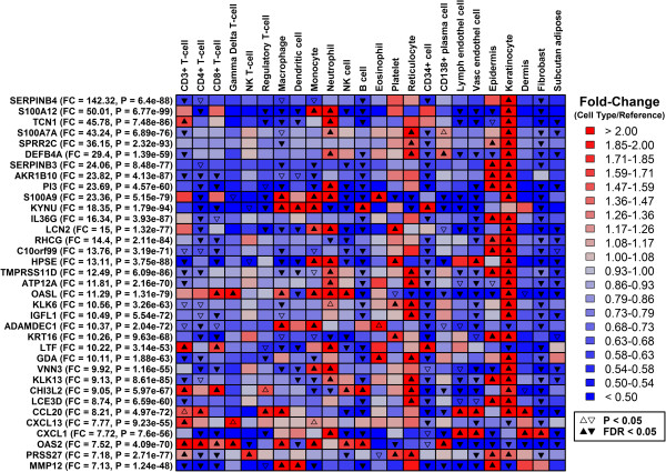
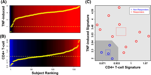
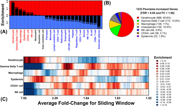
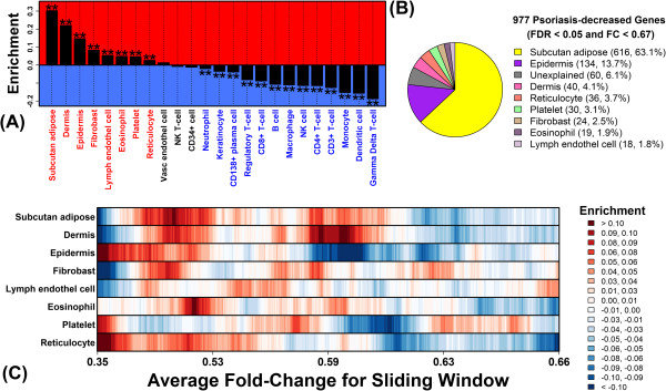
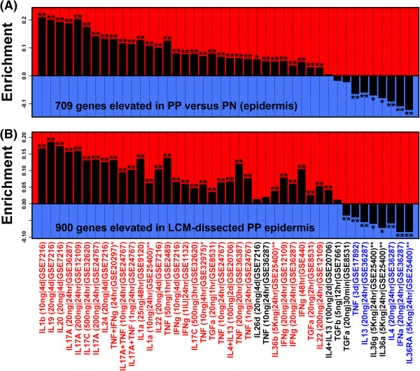
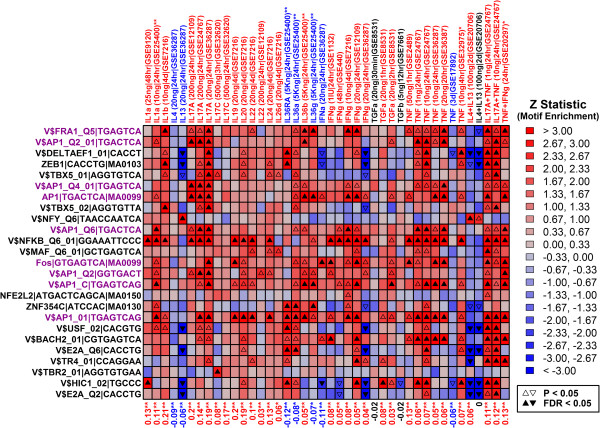
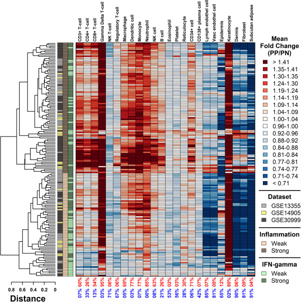
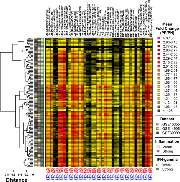
We identified 1233 psoriasis-increased DEGs and 977 psoriasis-decreased DEGs. Increased DEGs were attributed to keratinocyte activity (56%) and infiltration of lesions by T-cells (14%) and macrophages (11%). Decreased DEGs, in contrast, were associated with adipose tissue (63%), epidermis (14%) and dermis (4%). KC/epidermis DEGs were enriched for genes induced by IL-1, IL-17A and IL-20 family cytokines, and were also disproportionately associated with AP-1 binding sites. Among all patients, 50% exhibited a heightened inflammatory signature, with increased expression of genes expressed by T-cells, monocytes and dendritic cells. 66% of patients displayed an IFN-γ-strong signature, with increased expression of genes induced by IFN-γ in addition to several other cytokines (e.g., IL-1, IL-17A and TNF). We show that such differences in gene expression can be used to differentiate between etanercept responders and non-responders.
Psoriasis DEGs are partly explained by shifts in the cellular composition of psoriasis lesions. Epidermal DEGs, however, may be driven by the activity of AP-1 and cellular responses to IL-1, IL-17A and IL-20 family cytokines. Among patients, we uncovered a range of inflammatory- and cytokine-associated gene expression patterns. Such patterns may provide biomarkers for predicting individual responses to biologic therapy.
银屑病皮损的特征是基因表达的大规模改变。然而,导致差异表达基因(DEGs)的机制尚不完全清楚。我们分析了现有的数据集,以评估来自 163 名银屑病患者皮损的全基因组表达。我们的目的是确定驱动差异表达的机制,并在这个大样本中描述皮损之间的异质性。
我们鉴定出了 1233 个银屑病增加的 DEGs 和 977 个银屑病减少的 DEGs。增加的 DEGs归因于角质形成细胞活性(56%)、T 细胞(14%)和巨噬细胞(11%)浸润皮损。相比之下,减少的 DEGs与脂肪组织(63%)、表皮(14%)和真皮(4%)有关。KC/表皮 DEGs 富含由 IL-1、IL-17A 和 IL-20 家族细胞因子诱导的基因,并且与 AP-1 结合位点不成比例地相关。在所有患者中,有 50%表现出强烈的炎症特征,T 细胞、单核细胞和树突状细胞表达的基因表达增加。66%的患者表现出 IFN-γ 强特征,除了几种其他细胞因子(如 IL-1、IL-17A 和 TNF)外,还诱导 IFN-γ 诱导的基因表达。我们表明,基因表达的这些差异可用于区分依那西普的应答者和非应答者。
银屑病 DEGs 部分可由银屑病皮损中细胞组成的变化来解释。然而,表皮 DEGs 可能由 AP-1 的活性以及角质形成细胞对 IL-1、IL-17A 和 IL-20 家族细胞因子的反应驱动。在患者中,我们发现了一系列炎症和细胞因子相关的基因表达模式。这些模式可能为预测个体对生物治疗的反应提供生物标志物。