• 文献检索
  • 文档翻译
  • 深度研究
  • 学术资讯
  • Suppr Zotero 插件Zotero 插件
  • 邀请有礼
  • 套餐&价格
  • 历史记录
应用&插件
Suppr Zotero 插件Zotero 插件浏览器插件Mac 客户端Windows 客户端微信小程序
定价
高级版会员购买积分包购买API积分包
服务
文献检索文档翻译深度研究API 文档MCP 服务
关于我们
关于 Suppr公司介绍联系我们用户协议隐私条款
关注我们

Suppr 超能文献

核心技术专利:CN118964589B侵权必究
粤ICP备2023148730 号-1Suppr @ 2026

文献检索

告别复杂PubMed语法,用中文像聊天一样搜索,搜遍4000万医学文献。AI智能推荐,让科研检索更轻松。

立即免费搜索

文件翻译

保留排版,准确专业,支持PDF/Word/PPT等文件格式,支持 12+语言互译。

免费翻译文档

深度研究

AI帮你快速写综述,25分钟生成高质量综述,智能提取关键信息,辅助科研写作。

立即免费体验

圆锥角膜的影像学检查方法。

Imaging modalities in keratoconus.

机构信息

Department of Cornea and Refractive Surgery, Narayana Nethralaya Superspeciality Eye Hospital and Post Graduate Institute of Ophthalmology, Bangalore, Karnataka, India.

出版信息

Indian J Ophthalmol. 2013 Aug;61(8):394-400. doi: 10.4103/0301-4738.116058.

DOI:10.4103/0301-4738.116058
PMID:23925322
原文链接:https://pmc.ncbi.nlm.nih.gov/articles/PMC3775072/
Abstract

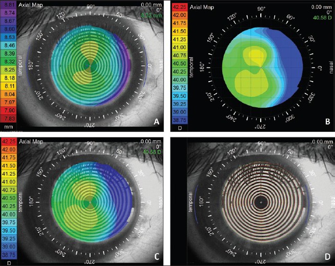
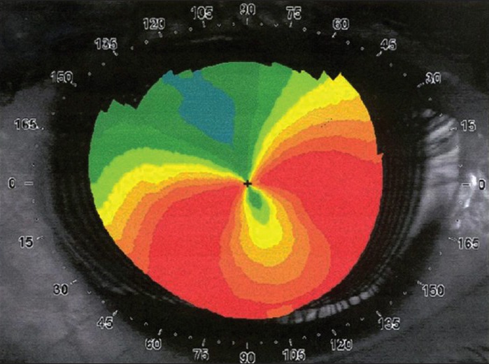
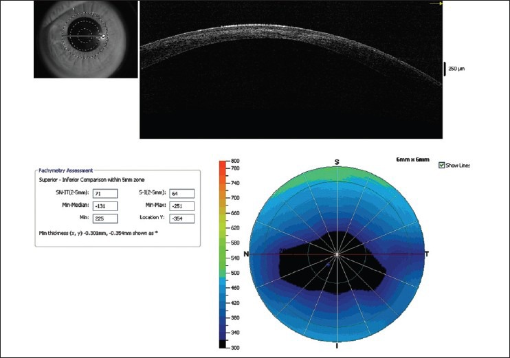
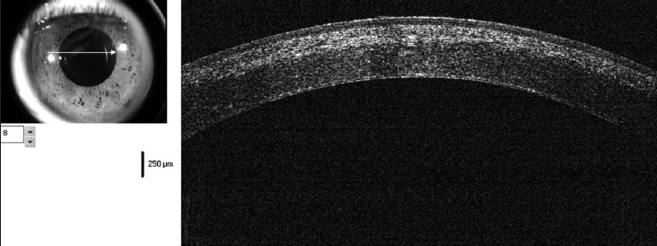
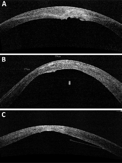
Diagnosis of keratoconus has greatly improved from simple clinical diagnosis with the advent of better diagnostic devices like corneal topographers based on placido disc, elevation based topographers and lately optical coherence tomography (OCT). These instruments are quite sensitive to pick up early keratoconus, which could help refractive surgeons to avoid serious complications like ectasia following keratorefractive surgeries. Each of these instruments has their advantages and disadvantages; in spite of that each one of them has its own place in the clinical practice. Currently, placido disc based topographers are the most commonly used topographers all over the world. There are many different companies making such devices, which follow the different techniques and color for the display. Due to these differences they are not directly comparable to each other. Various quantitative indices based on these topographers have been suggested and validated by different authors to aid in the diagnosis and quantification of keratoconus. OCT with its higher resolution and deeper penetration has created its place in the diagnostic armamentarium for keratoconus.

摘要

圆锥角膜的诊断随着更好的诊断设备的出现(如基于角膜面镜的角膜地形图仪、基于高度计的角膜地形图仪以及最近的光学相干断层扫描 (OCT))而得到了极大的改善。这些仪器对早期圆锥角膜的检测非常敏感,可以帮助屈光外科医生避免角膜屈光手术后出现像扩张这样的严重并发症。这些仪器中的每一种都有其优点和缺点;尽管如此,它们中的每一种都在临床实践中有其自己的地位。目前,基于角膜面镜的角膜地形图仪是全世界最常用的角膜地形图仪。有许多不同的公司制造这种设备,它们采用不同的技术和颜色进行显示。由于这些差异,它们彼此之间无法直接进行比较。不同的作者提出了基于这些角膜地形图仪的各种定量指标,并对其进行了验证,以帮助诊断和量化圆锥角膜。OCT 具有更高的分辨率和更深的穿透性,在圆锥角膜的诊断工具中占据了一席之地。

https://cdn.ncbi.nlm.nih.gov/pmc/blobs/39d3/3775072/51e6fd2d9825/IJO-61-394-g012.jpg
https://cdn.ncbi.nlm.nih.gov/pmc/blobs/39d3/3775072/7b062ab44138/IJO-61-394-g001.jpg
https://cdn.ncbi.nlm.nih.gov/pmc/blobs/39d3/3775072/e07cb9aa9e59/IJO-61-394-g002.jpg
https://cdn.ncbi.nlm.nih.gov/pmc/blobs/39d3/3775072/ac93030489c6/IJO-61-394-g004.jpg
https://cdn.ncbi.nlm.nih.gov/pmc/blobs/39d3/3775072/cac544b22984/IJO-61-394-g007.jpg
https://cdn.ncbi.nlm.nih.gov/pmc/blobs/39d3/3775072/c42f17e5ab0d/IJO-61-394-g008.jpg
https://cdn.ncbi.nlm.nih.gov/pmc/blobs/39d3/3775072/bd158e685e1f/IJO-61-394-g009.jpg
https://cdn.ncbi.nlm.nih.gov/pmc/blobs/39d3/3775072/b504f6bbe9cd/IJO-61-394-g010.jpg
https://cdn.ncbi.nlm.nih.gov/pmc/blobs/39d3/3775072/04336a167fe7/IJO-61-394-g011.jpg
https://cdn.ncbi.nlm.nih.gov/pmc/blobs/39d3/3775072/51e6fd2d9825/IJO-61-394-g012.jpg

相似文献

1
Imaging modalities in keratoconus.圆锥角膜的影像学检查方法。
Indian J Ophthalmol. 2013 Aug;61(8):394-400. doi: 10.4103/0301-4738.116058.
2
Cornea and anterior eye assessment with placido-disc keratoscopy, slit scanning evaluation topography and scheimpflug imaging tomography.角膜和眼前节评估的角膜地形图、扫描检眼镜评估地形和共焦激光角膜显微镜成像断层扫描。
Indian J Ophthalmol. 2018 Mar;66(3):360-366. doi: 10.4103/ijo.IJO_850_17.
3
Comparison of corneal measurements in keratoconus using swept-source optical coherence tomography and combined Placido-Scheimpflug imaging.使用扫频源光学相干断层扫描和普拉西多-谢姆弗鲁格联合成像技术对比圆锥角膜的角膜测量结果
Acta Ophthalmol. 2017 Sep;95(6):e486-e494. doi: 10.1111/aos.13298. Epub 2016 Nov 2.
4
[New diagnostic methods for imaging the anterior segment of the eye to enable treatment modalities selection].[用于眼部前段成像以辅助治疗方式选择的新诊断方法]
Nippon Ganka Gakkai Zasshi. 2011 Mar;115(3):297-322; discussion 323.
5
Keratoconus diagnosis with optical coherence tomography pachymetry mapping.基于光学相干断层扫描测厚成像的圆锥角膜诊断
Ophthalmology. 2008 Dec;115(12):2159-66. doi: 10.1016/j.ophtha.2008.08.004. Epub 2008 Nov 5.
6
Comparison of three-dimensional optical coherence tomography and combining a rotating Scheimpflug camera with a Placido topography system for forme fruste keratoconus diagnosis.三种光学相干断层扫描技术与旋转 Scheimpflug 相机联合 Placido 角膜地形图系统在诊断亚临床圆锥角膜中的比较。
Br J Ophthalmol. 2013 Dec;97(12):1554-9. doi: 10.1136/bjophthalmol-2013-303477. Epub 2013 Sep 30.
7
Scheimpflug imaging for keratoconus and ectatic disease.Scheimpflug 成像在圆锥角膜和扩张性疾病中的应用。
Indian J Ophthalmol. 2013 Aug;61(8):401-6. doi: 10.4103/0301-4738.116059.
8
Characterization of corneal structure in keratoconus.圆锥角膜的角膜结构特征。
J Cataract Refract Surg. 2012 Dec;38(12):2167-83. doi: 10.1016/j.jcrs.2012.10.022.
9
The Evolution of Diagnostics for Keratoconus: From Ophthalmometry to Biomechanics.圆锥角膜诊断技术的演进:从眼压计到生物力学。
Semin Ophthalmol. 2023 Apr;38(3):265-274. doi: 10.1080/08820538.2022.2152716. Epub 2023 Jan 4.
10
A review of imaging modalities for detecting early keratoconus.早期圆锥角膜的影像学检测方法综述。
Eye (Lond). 2021 Jan;35(1):173-187. doi: 10.1038/s41433-020-1039-1. Epub 2020 Jul 16.

引用本文的文献

1
Corneal Topography - a Review of Available Investigation Methods and Impact in the Diagnosis and Follow-Up of Keratoconus.角膜地形图——现有检查方法综述及其在圆锥角膜诊断和随访中的作用
Maedica (Bucur). 2025 Jun;20(2):374-382. doi: 10.26574/maedica.2025.20.2.374.
2
Strategies for Early Keratoconus Diagnosis: A Narrative Review of Evaluating Affordable and Effective Detection Techniques.早期圆锥角膜诊断策略:关于评估经济有效检测技术的叙述性综述
J Clin Med. 2025 Jan 13;14(2):460. doi: 10.3390/jcm14020460.
3
The role of corneal epithelial thickness map in detecting early keratoconus.

本文引用的文献

1
Corneal epithelial thickness mapping by Fourier-domain optical coherence tomography in normal and keratoconic eyes.傅里叶域光学相干断层扫描对正常眼和圆锥角膜眼的角膜上皮厚度测绘。
Ophthalmology. 2012 Dec;119(12):2425-33. doi: 10.1016/j.ophtha.2012.06.023. Epub 2012 Aug 20.
2
Revisiting corneal topography for the diagnosis of keratoconus: use of Rabinowitz's KISA% index.重新审视角膜地形图在圆锥角膜诊断中的应用:Rabinowitz的KISA%指数的使用
Clin Ophthalmol. 2012;6:181-4. doi: 10.2147/OPTH.S24219. Epub 2012 Jan 26.
3
Pachymetric mapping with Fourier-domain optical coherence tomography.
角膜上皮厚度图在早期圆锥角膜检测中的作用。
Graefes Arch Clin Exp Ophthalmol. 2025 Apr;263(4):1035-1044. doi: 10.1007/s00417-024-06682-9. Epub 2024 Nov 13.
4
Inflammatory and genomic interactions within keratoconus susceptible patients: a nationwide registered case-control study.圆锥角膜易感患者的炎症与基因组相互作用:一项全国性注册病例对照研究。
Eye Vis (Lond). 2024 Oct 2;11(1):40. doi: 10.1186/s40662-024-00407-z.
5
Incidence and prevalence of keratoconus in Taiwan during 2000-2018 and their association with the use of corneal topography and tomography.2000年至2018年台湾圆锥角膜的发病率和患病率及其与角膜地形图和断层扫描使用的关联。
Eye (Lond). 2024 Mar;38(4):745-751. doi: 10.1038/s41433-023-02767-7. Epub 2023 Oct 19.
6
Association of 2 Lysyl Oxidase Gene Single Nucleotide Polymorphisms with Keratoconus: A Nationwide Registration Study.赖氨酰氧化酶基因单核苷酸多态性与圆锥角膜的关联:一项全国性登记研究
Ophthalmol Sci. 2022 Nov 9;3(2):100247. doi: 10.1016/j.xops.2022.100247. eCollection 2023 Jun.
7
Topographic changes in contralateral eye of a case of Terrien's marginal degeneration.一例边缘性角膜变性患者对侧眼的地形变化。
BMJ Case Rep. 2022 Jul 4;15(7):e249493. doi: 10.1136/bcr-2022-249493.
8
Biometric Determinants of Epithelial Thickness Profile Across a Wide Range of Refractive Errors.不同屈光不正情况下上皮厚度分布的生物特征决定因素
Ophthalmol Ther. 2022 Jun;11(3):1089-1100. doi: 10.1007/s40123-022-00489-9. Epub 2022 Mar 14.
9
Surgically induced astigmatism and posterior corneal curvature changes following phacoemulsification.超声乳化白内障吸除术后手术源性散光及后角膜曲率变化。
Indian J Ophthalmol. 2022 Feb;70(2):406-412. doi: 10.4103/ijo.IJO_882_21.
10
Sequence of events leading to diagnosis of keratoconus and its impact on quality of life.导致圆锥角膜诊断的事件序列及其对生活质量的影响。
Indian J Ophthalmol. 2021 Dec;69(12):3478-3481. doi: 10.4103/ijo.IJO_399_21.
应用傅里叶域光学相干断层成像技术进行角膜生物力学测量。
J Cataract Refract Surg. 2010 May;36(5):826-31. doi: 10.1016/j.jcrs.2009.11.016.
4
Use of anterior segment optical coherence tomography to study corneal changes after collagen cross-linking.使用眼前节光学相干断层扫描技术研究胶原交联术后的角膜变化。
Am J Ophthalmol. 2009 Dec;148(6):844-51.e2. doi: 10.1016/j.ajo.2009.06.031. Epub 2009 Sep 24.
5
Keratoconus: classification scheme based on videokeratography and clinical signs.圆锥角膜:基于角膜地形图和临床体征的分类方案。
J Cataract Refract Surg. 2009 Sep;35(9):1597-603. doi: 10.1016/j.jcrs.2009.03.050.
6
Corneal epithelial thickness profile in the diagnosis of keratoconus.圆锥角膜诊断中的角膜上皮厚度分布
J Refract Surg. 2009 Jul;25(7):604-10. doi: 10.3928/1081597X-20090610-06.
7
Keratoconus diagnosis with optical coherence tomography pachymetry mapping.基于光学相干断层扫描测厚成像的圆锥角膜诊断
Ophthalmology. 2008 Dec;115(12):2159-66. doi: 10.1016/j.ophtha.2008.08.004. Epub 2008 Nov 5.
8
CLMI: the cone location and magnitude index.CLMI:视锥位置与大小指数。
Cornea. 2008 May;27(4):480-7. doi: 10.1097/ICO.0b013e31816485d3.
9
Laser scanning in vivo confocal analysis of keratocyte density in keratoconus.圆锥角膜中角膜细胞密度的激光扫描活体共聚焦分析
Ophthalmology. 2008 May;115(5):845-50. doi: 10.1016/j.ophtha.2007.04.067. Epub 2007 Sep 6.
10
Treatment of progressive keratoconus by riboflavin-UVA-induced cross-linking of corneal collagen: ultrastructural analysis by Heidelberg Retinal Tomograph II in vivo confocal microscopy in humans.核黄素-紫外线A诱导角膜胶原交联治疗进行性圆锥角膜:应用海德堡视网膜断层扫描仪II及活体共聚焦显微镜进行超微结构分析(人体研究)
Cornea. 2007 May;26(4):390-7. doi: 10.1097/ICO.0b013e318030df5a.