Shanghai Key Laboratory of Orthopaedic Implants, Department of Orthopaedic Surgery, Shanghai Ninth People's Hospital, Shanghai Jiaotong University School of Medicine, Shanghai, People's Republic of China.
Int J Nanomedicine. 2013;8:3093-105. doi: 10.2147/IJN.S48084. Epub 2013 Aug 14.
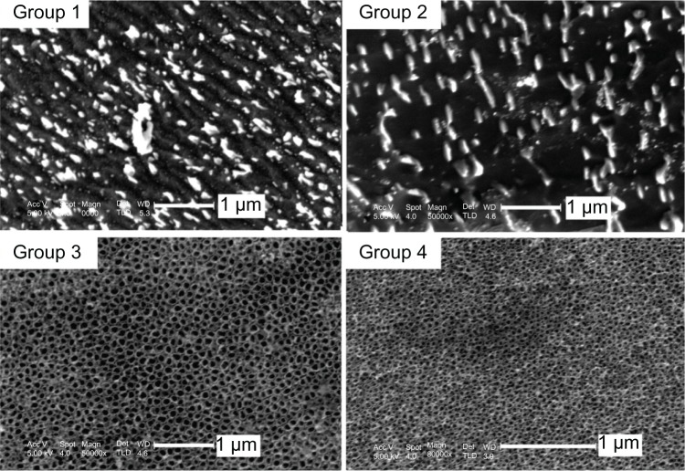
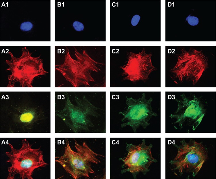
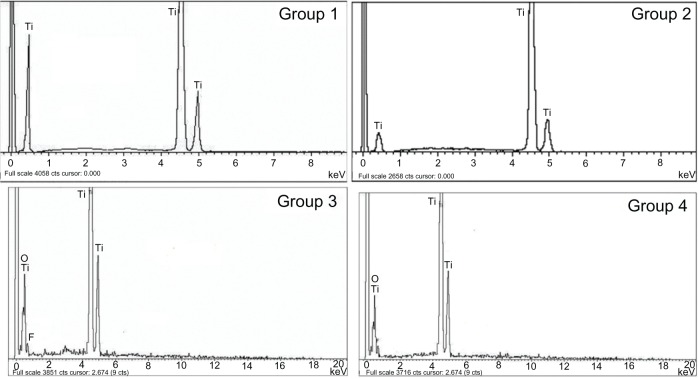
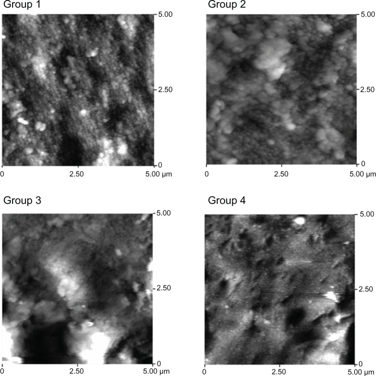
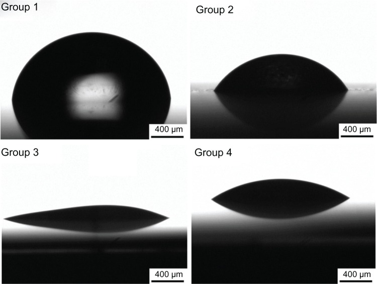
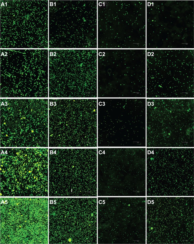
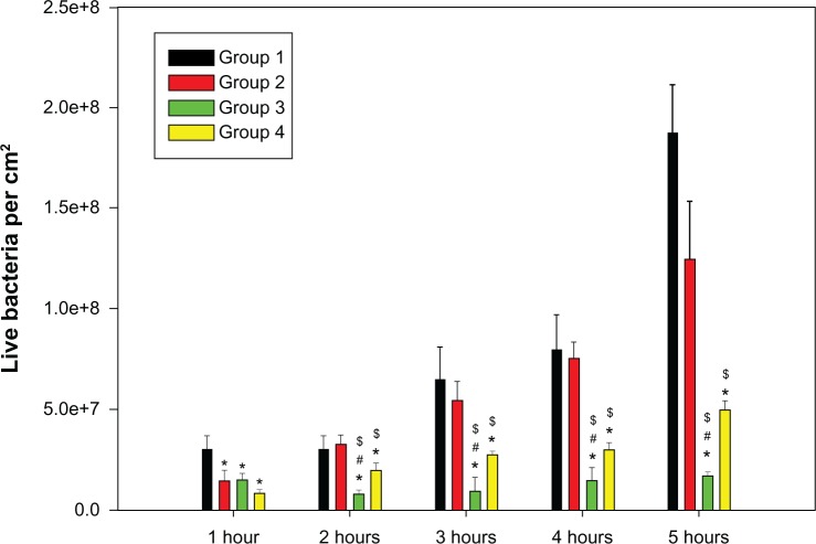
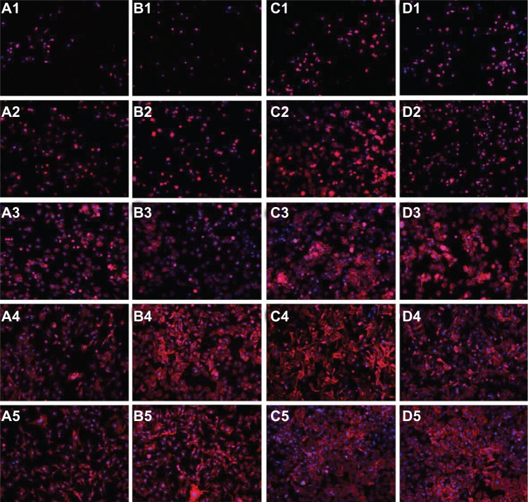
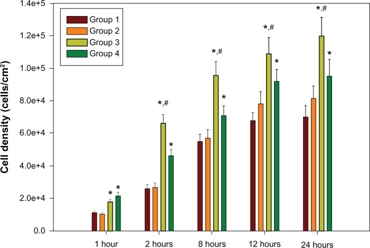
Competition occurs between the osteoblasts in regional microenvironments and pathogens introduced during surgery, on the surface of bone implants, such as joint prostheses. The aim of this study was to modulate bacterial and osteoblast adhesion on implant surfaces by using a nanotube array. Titanium oxide (TiO2) nanotube arrays, 30 nm or 80 nm in diameter, were prepared by a two-step anodization on titanium substrates. Mechanically polished and acid-etched titanium samples were also prepared to serve as control groups. The standard strains of Staphylococcus epidermidis (S. epidermidis, American Type Culture Collection [ATCC]35984) and mouse C3H10T1/2 cell lines with osteogenic potential were used to evaluate the different responses to the nanotube arrays, in bacteria and eukaryotic cells. We found that the initial adhesion and colonization of S. epidermidis on the surface of the TiO2 nanotube arrays were significantly reduced and that the adhesion of C3H10T1/2 cells on the surface of the TiO2 nanotube arrays was significantly enhanced when compared with the control samples. Based on a surface analysis of all four groups, we observed increased surface roughness, decreased water contact angles, and an enhanced concentration of oxygen and fluorine atoms on the TiO2 nanotube surface. We conclude that the TiO2 nanotube surface can reduce bacterial colonization and enhance C3H10T1/2 cell adhesion; multiple physical and chemical properties of the TiO2 nanotube surface may contribute to these dual effects.
在骨植入物表面(如关节假体)的局部微环境中,成骨细胞与手术中引入的病原体之间存在竞争。本研究旨在通过使用纳米管阵列来调节细菌和成骨细胞在植入物表面的黏附。通过两步阳极氧化在钛基底上制备了直径为 30nm 或 80nm 的氧化钛(TiO2)纳米管阵列。还制备了机械抛光和酸蚀的钛样品作为对照组。使用标准表皮葡萄球菌(S. epidermidis,美国典型培养物保藏中心 [ATCC]35984)菌株和具有成骨潜能的小鼠 C3H10T1/2 细胞系来评估纳米管阵列对细菌和真核细胞的不同反应。我们发现,与对照样品相比,S. epidermidis 在 TiO2 纳米管阵列表面的初始黏附和定植明显减少,而 C3H10T1/2 细胞在 TiO2 纳米管阵列表面的黏附明显增强。基于对所有四个组的表面分析,我们观察到表面粗糙度增加,水接触角减小,以及 TiO2 纳米管表面的氧和氟原子浓度增强。我们得出结论,TiO2 纳米管表面可以减少细菌定植并增强 C3H10T1/2 细胞黏附;TiO2 纳米管表面的多种物理和化学性质可能促成了这两种效应。