Department of Pharmacy, Namur Research Institute for LIfe Sciences, Namur Thrombosis and Hemostasis Center (NTHC), University of Namur, Belgium ; Haematology Laboratory, Namur Research Institute for LIfe Sciences, Namur Thrombosis and Hemostasis Center (NTHC), CHU Mont-Godinne, Université Catholique de Louvain, Belgium.
J Extracell Vesicles. 2013 Mar 18;2. doi: 10.3402/jev.v2i0.19728. eCollection 2013.
Patients with cancer have a 7- to 10-fold increased risk of developing venous thromboembolism. Circulating microvesicles could be a useful predictive biomarker for venous thromboembolism in cancer. Validated and standardised techniques that could be used to determine the complete microvesicle phenotype are required.
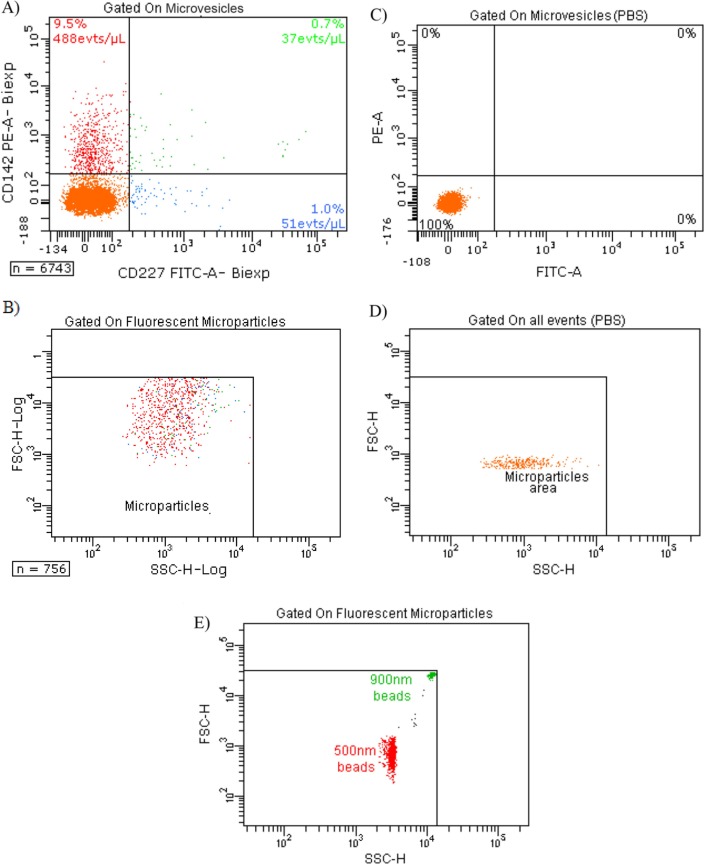
These were two-fold: a) to characterise tissue factor (TF)-bearing microvesicles released by cultured breast cancer cells MDA-MB-231 by flow cytometry (FCM), transmission electron microscopy (TEM) and thrombin generation assay (TGA); and b) to validate the sensitivity and variability intra/inter-assay of TGA as a useful method to study the procoagulant activity (PCA) of microvesicles.
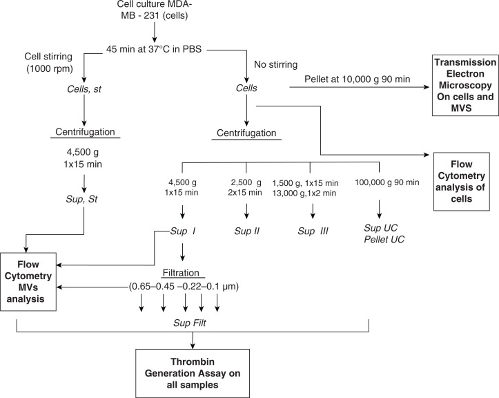
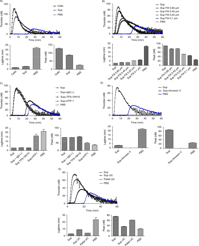
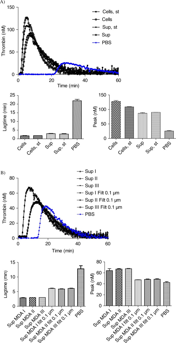
Cultured breast cancer cells MDA-MB-231 were incubated for 45 minutes at 37°C. Samples were then centrifuged or not at 4,500 g for 15 minutes, and cells and MVs or MV-containing supernatants were used for TEM, FCM and TGA. In activity assays, microvesicles (i.e. cell-depleted supernatants) were incubated with anti-TF antibodies or with annexin V to assess the contribution of TF and phospholipids to the PCA. Alternatively, supernatants were filtered through 0.1, 0.22, 0.45 or 0.65 µm membranes and subjected to TGA.
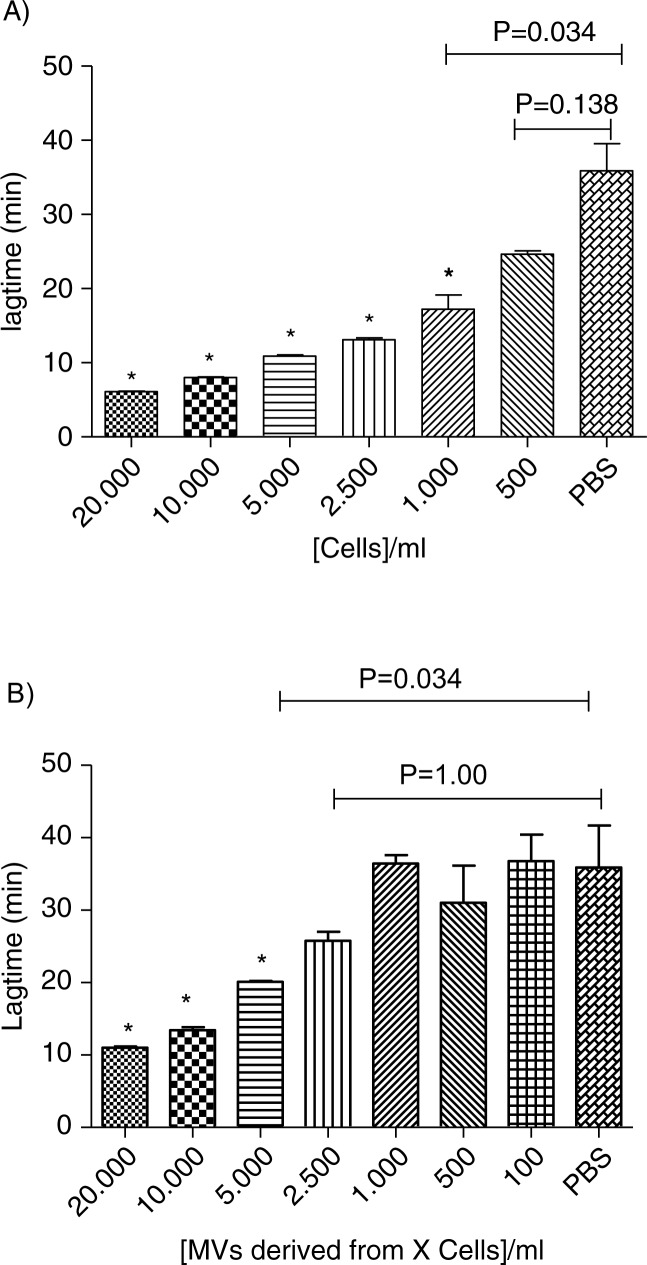
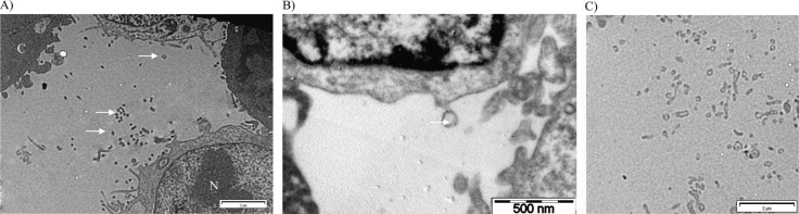
The majority of the PCA was associated with microvesicles smaller than 0.1 µm, and the mean microvesicle size estimated by TEM after 10,000 g centrifugation was 121±54 nm with a majority of vesicles between 100 and 200 nm. Microvesicles derived from 5,000 MDA-MB-231cells/ml were sufficient to significantly increase the thrombin generation of normal pooled plasma.
TEM, FCM and filtration coupled to TGA represent a useful combination to study the PCA of TF-bearing microvesicles, whatever their size. And it will be interesting to implement these techniques in patients.
癌症患者发生静脉血栓栓塞的风险增加了 7 至 10 倍。循环中的微囊泡可能是癌症患者静脉血栓栓塞的有用预测生物标志物。需要验证和标准化可用于确定完整微囊泡表型的技术。
有两个目标:a)通过流式细胞术(FCM)、透射电子显微镜(TEM)和凝血酶生成试验(TGA)描述培养的乳腺癌细胞 MDA-MB-231 释放的组织因子(TF)携带的微囊泡;b)验证 TGA 作为研究微囊泡促凝活性(PCA)的有用方法的灵敏度和内/间试验变异性。
培养的乳腺癌细胞 MDA-MB-231 在 37°C 下孵育 45 分钟。然后将样品在 4500g 下离心 15 分钟,使用细胞和 MV 或含 MV 的上清液进行 TEM、FCM 和 TGA。在活性测定中,将微囊泡(即细胞耗尽的上清液)与抗 TF 抗体或与 annexin V 孵育,以评估 TF 和磷脂对 PCA 的贡献。或者,将上清液通过 0.1、0.22、0.45 或 0.65 µm 膜过滤,并进行 TGA。
大多数 PCA 与小于 0.1 µm 的微囊泡相关,TEM 估计的经 10000g 离心后的平均微囊泡大小为 121±54nm,大多数囊泡在 100 至 200nm 之间。来自 5000 个 MDA-MB-231 细胞/ml 的微囊泡足以显著增加正常混合血浆的凝血酶生成。
TEM、FCM 和与 TGA 结合的过滤代表了研究 TF 携带的微囊泡 PCA 的有用组合,无论其大小如何。在患者中实施这些技术将是有趣的。