Hamilton Glaucoma Center and Department of Ophthalmology, University of California, San Diego, La Jolla, California; Glaucoma Sector, Department of Ophthalmology, Geneva University Hospitals, Geneva, Switzerland.
Hamilton Glaucoma Center and Department of Ophthalmology, University of California, San Diego, La Jolla, California.
Ophthalmology. 2013 Dec;120(12):2508-2516. doi: 10.1016/j.ophtha.2013.07.040. Epub 2013 Sep 8.
To evaluate changes in peripapillary and macular choroidal thickness and volume after the water-drinking test (WDT) using swept-source optical coherence tomography (SS OCT).
Prospective, cross-sectional, observational study.
Fifty-six eyes of 28 healthy volunteers.
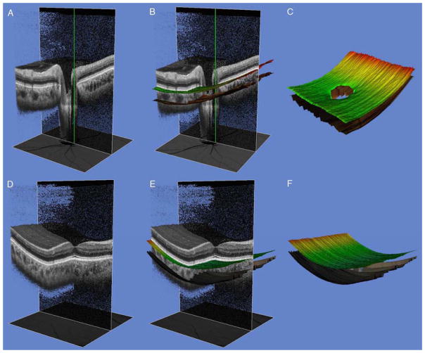
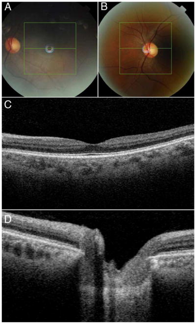
Participants underwent a 3-dimensional optic disc and macula scanning protocol with a prototype SS OCT (Topcon, Inc., Tokyo, Japan) at baseline and 15, 30, 45, and 120 minutes after the start of the WDT. The WDT consisted of drinking 1000 ml of water within 5 minutes. Objective measurements of the choroid were obtained with automated segmentation of the choroidal boundaries.
Choroidal thickness and volume.
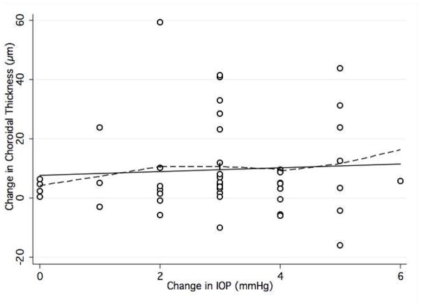
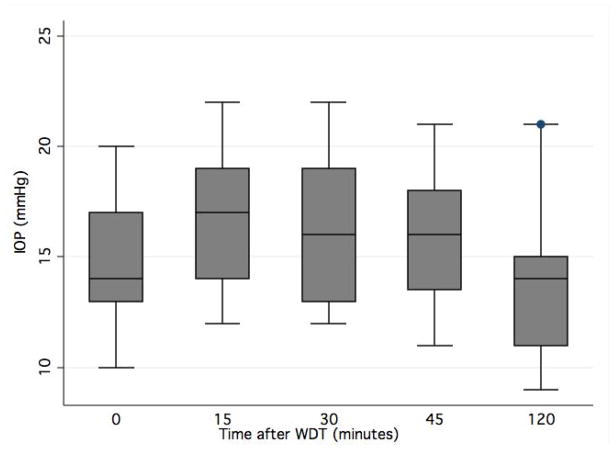
Mean age ± standard deviation of participants was 35.6 ± 9.1 years. Intraocular pressure (IOP) increased from 14.9 ± 2.7 mmHg at baseline to a peak of 16.8 ± 3.0 mmHg 15 minutes after the WDT (P < 0.001). Mean baseline choroidal thickness and volume were 181.3 ± 50.8 μm and 6.19 ± 1.80 mm(3), respectively, at the optic disc and 217.4 ± 43.6 μm and 7.83 ± 1.55 mm(3), respectively, at the macula. After the WDT, peripapillary and macular choroidal thickness increased by a maximum of 5.7% (P<0.001) and 4.3% (P<0.001), respectively. Choroidal volumes increased by 6.4% (P<0.001) and 3.9% (P<0.001), respectively. There was no association between change in IOP and peripapillary (P = 0.27) or macular (P = 0.09) choroidal thickness.
Using automated segmentation of SS OCT measurements, significant increases in choroidal thickness and volume are observed after the WDT in healthy subjects.
使用扫频源光学相干断层扫描(SS-OCT)评估饮水试验(WDT)后视盘和黄斑脉络膜厚度和体积的变化。
前瞻性、横断面、观察性研究。
28 名健康志愿者的 56 只眼。
参与者在基线和 WDT 开始后 15、30、45 和 120 分钟进行了 3 维视盘和黄斑扫描方案,使用原型 SS-OCT(Topcon,Inc.,东京,日本)。WDT 包括在 5 分钟内饮用 1000 毫升水。使用脉络膜边界的自动分割获得脉络膜的客观测量值。
脉络膜厚度和体积。
参与者的平均年龄±标准差为 35.6±9.1 岁。眼压(IOP)从基线时的 14.9±2.7mmHg 升高到 WDT 后 15 分钟时的 16.8±3.0mmHg(P<0.001)。视盘处的平均基线脉络膜厚度和体积分别为 181.3±50.8μm 和 6.19±1.80mm3,黄斑处分别为 217.4±43.6μm 和 7.83±1.55mm3。WDT 后,视盘和黄斑脉络膜厚度最大增加了 5.7%(P<0.001)和 4.3%(P<0.001)。脉络膜体积分别增加了 6.4%(P<0.001)和 3.9%(P<0.001)。IOP 的变化与视盘(P=0.27)或黄斑(P=0.09)脉络膜厚度之间无相关性。
在健康受试者中,使用 SS-OCT 测量的自动分割,WDT 后脉络膜厚度和体积明显增加。