Powers Thomas W, Jones E Ellen, Betesh Lucy R, Romano Patrick R, Gao Peng, Copland John A, Mehta Anand S, Drake Richard R
Department of Cell and Molecular Pharmacology and Experimental Therapeutics and MUSC Proteomics Center, Medical University of South Carolina , 173 Ashley Avenue, Charleston, South Carolina, 29425 United States.
Anal Chem. 2013 Oct 15;85(20):9799-806. doi: 10.1021/ac402108x. Epub 2013 Oct 3.
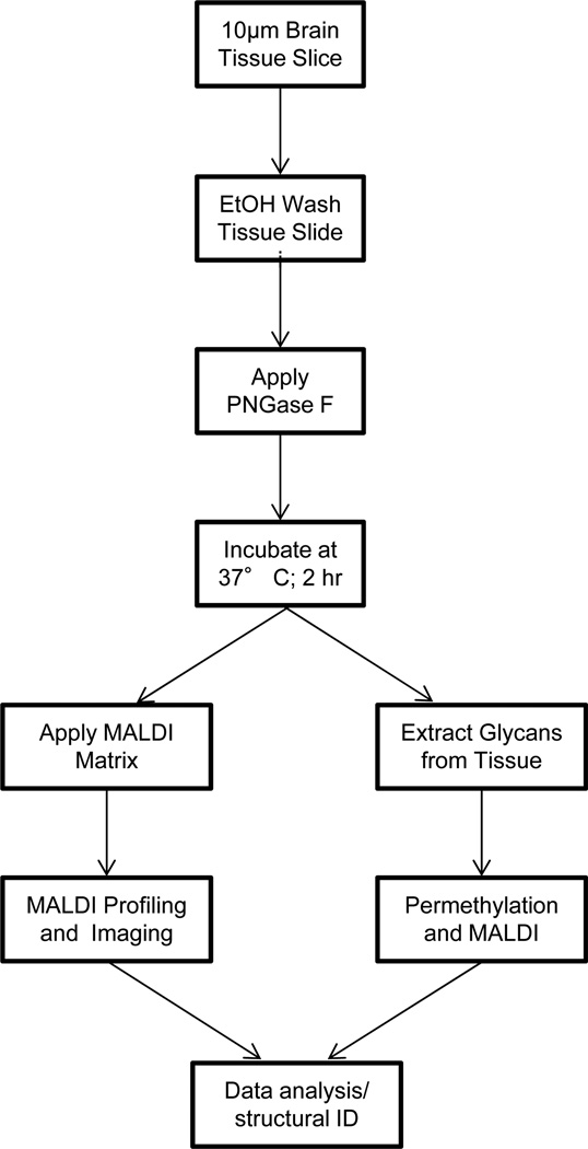
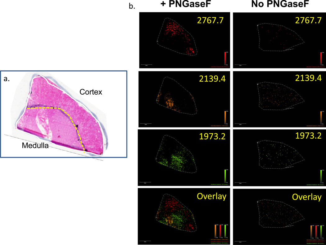
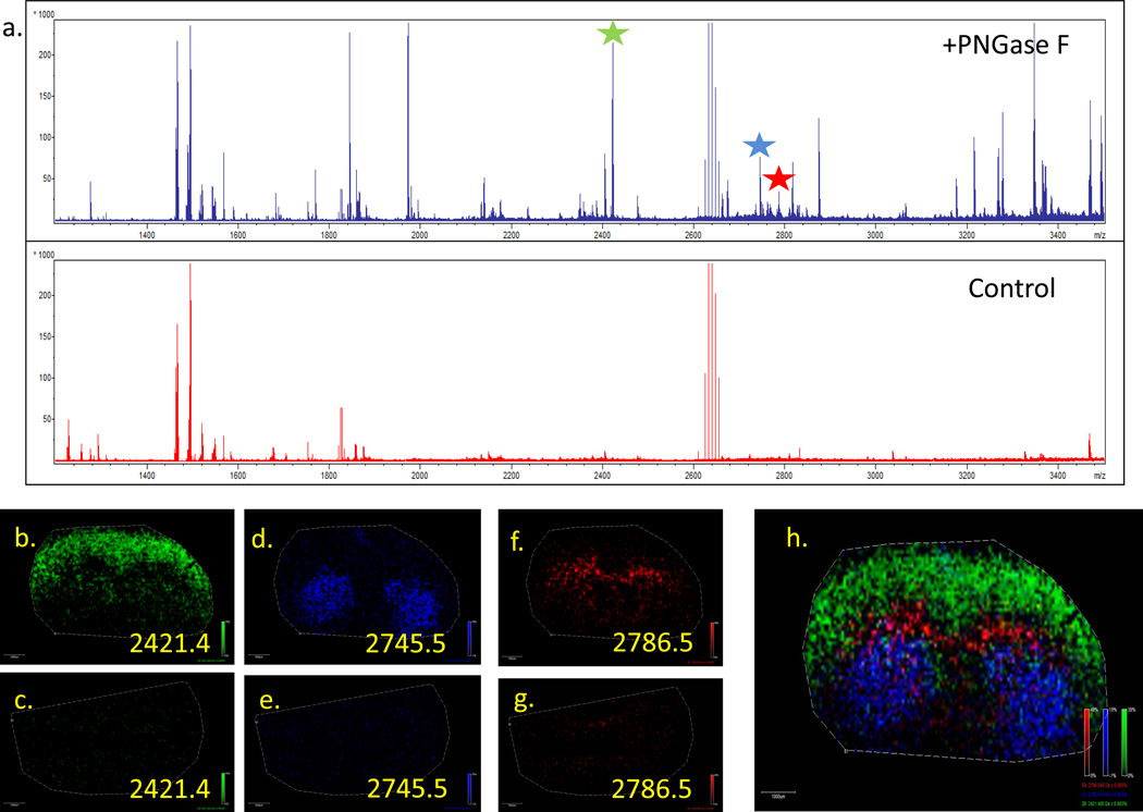
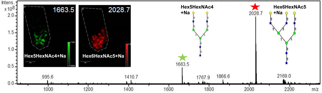
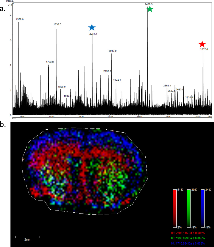
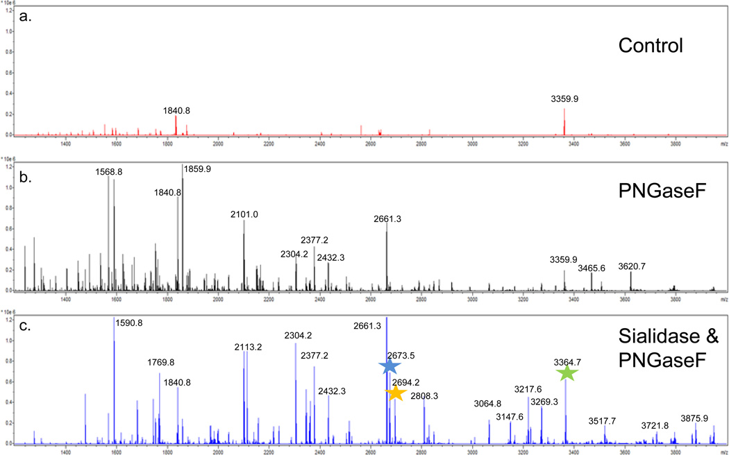
A new matrix assisted laser desorption ionization imaging mass spectrometry (MALDI-IMS) method to spatially profile the location and distribution of multiple N-linked glycan species in tissues is described. Application of an endoglycosidase, peptide N-glycosidase F (PNGaseF), directly on tissues followed by incubation releases N-linked glycan species amenable to detection by MALDI-IMS. The method has been designed to simultaneously profile the multiple glycan species released from intracellular organelle and cell surface glycoproteins, while maintaining histopathology compatible preparation workflows. A recombinant PNGaseF enzyme was sprayed uniformly across mouse brain tissue slides, incubated for 2 h, then sprayed with 2,5-dihydroxybenzoic acid matrix for MALDI-IMS analysis. Using this basic approach, global snapshots of major cellular N-linked glycoforms were detected, including their tissue localization and distribution, structure, and relative abundance. Off-tissue extraction and modification of glycans from similarly processed tissues and further mass spectrometry or HPLC analysis was done to assign structural designations. MALDI-IMS has primarily been utilized to spatially profile proteins, lipids, drug, and small molecule metabolites in tissues, but it has not been previously applied to N-linked glycan analysis. The translatable MALDI-IMS glycan profiling workflow described herein can readily be applied to any tissue type of interest. From a clinical diagnostics perspective, the ability to differentially profile N-glycans and correlate their molecular expression to histopathological changes can offer new approaches to identifying novel disease related targets for biomarker and therapeutic applications.
本文描述了一种新的基质辅助激光解吸电离成像质谱(MALDI-IMS)方法,用于在组织中对多种N-连接聚糖种类的位置和分布进行空间分析。将内切糖苷酶肽N-糖苷酶F(PNGaseF)直接应用于组织,随后进行孵育,可释放出适合通过MALDI-IMS检测的N-连接聚糖种类。该方法旨在同时分析从细胞内细胞器和细胞表面糖蛋白释放的多种聚糖种类,同时保持与组织病理学兼容的制备工作流程。将重组PNGaseF酶均匀喷洒在小鼠脑组织切片上,孵育2小时,然后喷洒2,5-二羟基苯甲酸基质用于MALDI-IMS分析。使用这种基本方法,检测到了主要细胞N-连接糖型的全局快照,包括它们在组织中的定位和分布、结构以及相对丰度。对经过类似处理的组织进行脱组织聚糖提取和修饰,并进一步进行质谱或HPLC分析,以确定结构命名。MALDI-IMS主要用于在组织中对蛋白质、脂质、药物和小分子代谢物进行空间分析,但此前尚未应用于N-连接聚糖分析。本文所述的可转化的MALDI-IMS聚糖分析工作流程可轻松应用于任何感兴趣的组织类型。从临床诊断的角度来看,对N-聚糖进行差异分析并将其分子表达与组织病理学变化相关联的能力,可以为识别用于生物标志物和治疗应用的新型疾病相关靶点提供新方法。