• 文献检索
  • 文档翻译
  • 深度研究
  • 学术资讯
  • Suppr Zotero 插件Zotero 插件
  • 邀请有礼
  • 套餐&价格
  • 历史记录
应用&插件
Suppr Zotero 插件Zotero 插件浏览器插件Mac 客户端Windows 客户端微信小程序
定价
高级版会员购买积分包购买API积分包
服务
文献检索文档翻译深度研究API 文档MCP 服务
关于我们
关于 Suppr公司介绍联系我们用户协议隐私条款
关注我们

Suppr 超能文献

核心技术专利:CN118964589B侵权必究
粤ICP备2023148730 号-1Suppr @ 2026

文献检索

告别复杂PubMed语法,用中文像聊天一样搜索,搜遍4000万医学文献。AI智能推荐,让科研检索更轻松。

立即免费搜索

文件翻译

保留排版,准确专业,支持PDF/Word/PPT等文件格式,支持 12+语言互译。

免费翻译文档

深度研究

AI帮你快速写综述,25分钟生成高质量综述,智能提取关键信息,辅助科研写作。

立即免费体验

头部和颈部磁共振成像扫描生成放射治疗用合成 CT 模型的方法研究。

Investigation of a method for generating synthetic CT models from MRI scans of the head and neck for radiation therapy.

机构信息

Department of Radiation Oncology, University of Michigan, Ann Arbor, MI 48109, USA.

出版信息

Phys Med Biol. 2013 Dec 7;58(23):8419-35. doi: 10.1088/0031-9155/58/23/8419. Epub 2013 Nov 11.

DOI:10.1088/0031-9155/58/23/8419
PMID:24217183
原文链接:https://pmc.ncbi.nlm.nih.gov/articles/PMC3886820/
Abstract

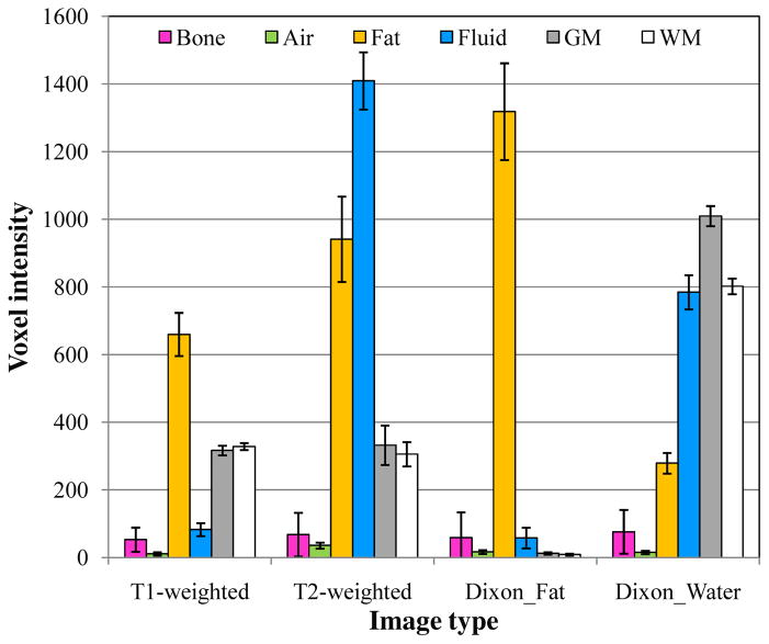
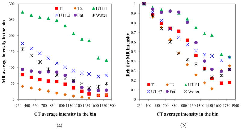
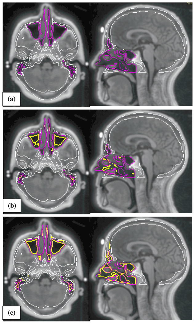
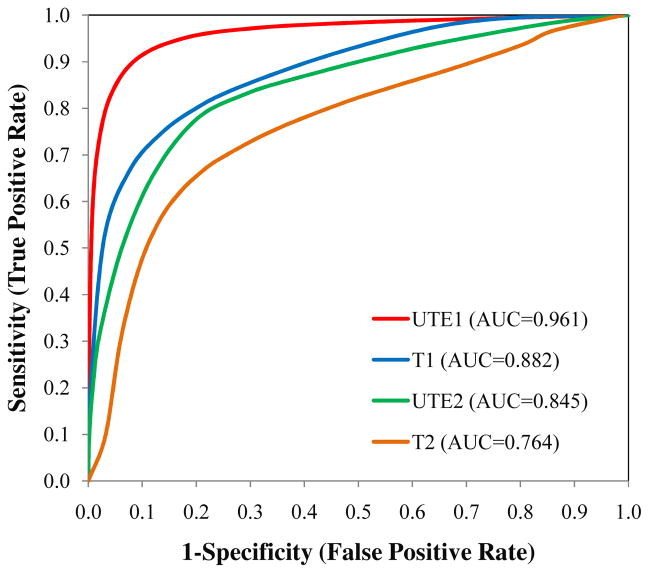
Magnetic resonance (MR) images often provide superior anatomic and functional information over computed tomography (CT) images, but generally are not used alone without CT images for radiotherapy treatment planning and image guidance. This study aims to investigate the potential of probabilistic classification of voxels from multiple MRI contrasts to generate synthetic CT ('MRCT') images. The method consists of (1) acquiring multiple MRI volumes: T1-weighted, T2-weighted, two echoes from a ultra-short echo time (UTE) sequence, and calculated fat and water image volumes using a Dixon method, (2) classifying tissues using fuzzy c-means clustering with a spatial constraint, (3) assigning attenuation properties with weights based on the probability of individual tissue classes being present in each voxel, and (4) generating a MRCT image volume from the sum of attenuation properties in each voxel. The capability of each MRI contrast to differentiate tissues of interest was investigated based on a retrospective analysis of ten patients. For one prospective patient, the correlation of skull intensities between CT and MR was investigated, the discriminatory power of MRI in separating air from bone was evaluated, and the generated MRCT image volume was qualitatively evaluated. Our analyses showed that one MRI volume was not sufficient to separate all tissue types, and T2-weighted images was more sensitive to bone density variation compared to other MRI image types. The short echo UTE image showed significant improvement in contrasting air versus bone, but could not completely separate air from bone without false labeling. Generated MRCT and CT images showed similar contrast between bone and soft/solid tissues. These results demonstrate the potential of the presented method to generate synthetic CT images to support the workflow of radiation oncology treatment planning and image guidance.

摘要

磁共振(MR)图像通常提供比计算机断层扫描(CT)图像更优越的解剖和功能信息,但通常不单独使用,而需要与 CT 图像结合,用于放射治疗计划和图像引导。本研究旨在探讨从多种 MRI 对比中对体素进行概率分类以生成合成 CT(“MRCT”)图像的潜力。该方法包括:(1)获取多个 MRI 容积:T1 加权、T2 加权、来自超短回波时间(UTE)序列的两个回波,以及使用 Dixon 方法计算脂肪和水图像容积;(2)使用具有空间约束的模糊 c-均值聚类对组织进行分类;(3)根据每个体素中存在的各个组织类别的概率为衰减特性分配权重;(4)根据每个体素中的衰减特性总和生成一个 MRCT 图像容积。基于对十名患者的回顾性分析,研究了每种 MRI 对比在区分感兴趣组织方面的能力。对于一名前瞻性患者,研究了 CT 和 MR 之间颅骨强度的相关性,评估了 MRI 在区分空气与骨骼方面的区分能力,并对生成的 MRCT 图像容积进行了定性评估。我们的分析表明,一个 MRI 容积不足以分离所有组织类型,与其他 MRI 图像类型相比,T2 加权图像对骨密度变化更敏感。短回波 UTE 图像在对比空气与骨骼方面显示出显著改善,但在没有虚假标记的情况下,无法完全分离空气与骨骼。生成的 MRCT 和 CT 图像显示出骨与软/固组织之间相似的对比度。这些结果表明,所提出的方法具有生成合成 CT 图像以支持放射肿瘤学治疗计划和图像引导工作流程的潜力。

https://cdn.ncbi.nlm.nih.gov/pmc/blobs/f745/3886820/cef1a1fd5873/nihms542406f11.jpg
https://cdn.ncbi.nlm.nih.gov/pmc/blobs/f745/3886820/04671931ac47/nihms542406f1.jpg
https://cdn.ncbi.nlm.nih.gov/pmc/blobs/f745/3886820/5245643f6acc/nihms542406f2.jpg
https://cdn.ncbi.nlm.nih.gov/pmc/blobs/f745/3886820/8f56c666dbc9/nihms542406f3.jpg
https://cdn.ncbi.nlm.nih.gov/pmc/blobs/f745/3886820/e2387acadc5a/nihms542406f4.jpg
https://cdn.ncbi.nlm.nih.gov/pmc/blobs/f745/3886820/9e0139e002f3/nihms542406f5.jpg
https://cdn.ncbi.nlm.nih.gov/pmc/blobs/f745/3886820/42fd9b46e18d/nihms542406f6.jpg
https://cdn.ncbi.nlm.nih.gov/pmc/blobs/f745/3886820/0d2e625ececf/nihms542406f7a.jpg
https://cdn.ncbi.nlm.nih.gov/pmc/blobs/f745/3886820/c60d9a90e4a6/nihms542406f8.jpg
https://cdn.ncbi.nlm.nih.gov/pmc/blobs/f745/3886820/f65d67fd8daf/nihms542406f9.jpg
https://cdn.ncbi.nlm.nih.gov/pmc/blobs/f745/3886820/53a13468acc6/nihms542406f10.jpg
https://cdn.ncbi.nlm.nih.gov/pmc/blobs/f745/3886820/cef1a1fd5873/nihms542406f11.jpg

相似文献

1
Investigation of a method for generating synthetic CT models from MRI scans of the head and neck for radiation therapy.头部和颈部磁共振成像扫描生成放射治疗用合成 CT 模型的方法研究。
Phys Med Biol. 2013 Dec 7;58(23):8419-35. doi: 10.1088/0031-9155/58/23/8419. Epub 2013 Nov 11.
2
Female pelvic synthetic CT generation based on joint intensity and shape analysis.基于联合强度和形状分析的女性盆腔合成CT生成
Phys Med Biol. 2017 Apr 21;62(8):2935-2949. doi: 10.1088/1361-6560/62/8/2935. Epub 2017 Mar 17.
3
Quantitative characterizations of ultrashort echo (UTE) images for supporting air-bone separation in the head.用于支持头部气骨分离的超短回波(UTE)图像的定量表征。
Phys Med Biol. 2015 Apr 7;60(7):2869-80. doi: 10.1088/0031-9155/60/7/2869. Epub 2015 Mar 17.
4
UTE-mDixon-based thorax synthetic CT generation.基于 UTE-mDixon 的胸部合成 CT 生成。
Med Phys. 2019 Aug;46(8):3520-3531. doi: 10.1002/mp.13574. Epub 2019 Jun 12.
5
MRI-based attenuation correction for hybrid PET/MRI systems: a 4-class tissue segmentation technique using a combined ultrashort-echo-time/Dixon MRI sequence.基于 MRI 的混合 PET/MRI 系统衰减校正:一种使用联合超短回波时间/Dixon MRI 序列的 4 类组织分割技术。
J Nucl Med. 2012 May;53(5):796-804. doi: 10.2967/jnumed.111.092577. Epub 2012 Apr 13.
6
MR-based attenuation correction for PET/MRI neurological studies with continuous-valued attenuation coefficients for bone through a conversion from R2* to CT-Hounsfield units.用于PET/MRI神经学研究的基于磁共振成像的衰减校正,通过从R2*转换为CT-亨氏单位来实现对具有连续值衰减系数的骨骼的校正。
Neuroimage. 2015 May 15;112:160-168. doi: 10.1016/j.neuroimage.2015.03.009. Epub 2015 Mar 14.
7
Magnetic Resonance-Based Automatic Air Segmentation for Generation of Synthetic Computed Tomography Scans in the Head Region.基于磁共振的自动空气分割技术,用于生成头部区域的合成计算机断层扫描。
Int J Radiat Oncol Biol Phys. 2015 Nov 1;93(3):497-506. doi: 10.1016/j.ijrobp.2015.07.001. Epub 2015 Jul 9.
8
A Technique to Rapidly Generate Synthetic Computed Tomography for Magnetic Resonance Imaging-Guided Online Adaptive Replanning: An Exploratory Study.一种用于快速生成磁共振成像引导在线自适应重计划的合成计算机断层扫描的技术:探索性研究。
Int J Radiat Oncol Biol Phys. 2019 Apr 1;103(5):1261-1270. doi: 10.1016/j.ijrobp.2018.12.008. Epub 2018 Dec 11.
9
Dosimetric evaluation of synthetic CT for head and neck radiotherapy generated by a patch-based three-dimensional convolutional neural network.基于补丁的三维卷积神经网络生成的头颈部放疗合成 CT 的剂量学评估。
Med Phys. 2019 Sep;46(9):4095-4104. doi: 10.1002/mp.13663. Epub 2019 Jul 9.
10
PET attenuation correction using synthetic CT from ultrashort echo-time MR imaging.使用来自超短回波时间磁共振成像的合成CT进行PET衰减校正。
J Nucl Med. 2014 Dec;55(12):2071-7. doi: 10.2967/jnumed.114.143958. Epub 2014 Nov 20.

引用本文的文献

1
Decision support using machine learning for predicting adequate bladder filling in prostate radiotherapy: a feasibility study.使用机器学习进行决策支持以预测前列腺放疗中膀胱的充分充盈:一项可行性研究。
Radiol Phys Technol. 2025 Jun 2. doi: 10.1007/s12194-025-00916-z.
2
Does dose calculation algorithm affect the dosimetric accuracy of synthetic CT for MR-only radiotherapy planning in brain tumors?剂量计算算法是否会影响脑肿瘤仅基于磁共振成像的放射治疗计划中合成CT的剂量学准确性?
J Appl Clin Med Phys. 2025 May;26(5):e70030. doi: 10.1002/acm2.70030. Epub 2025 Feb 19.
3
Advancements in synthetic CT generation from MRI: A review of techniques, and trends in radiation therapy planning.

本文引用的文献

1
Comparison of segmentation-based attenuation correction methods for PET/MRI: evaluation of bone and liver standardized uptake value with oncologic PET/CT data.基于分割的 PET/MRI 衰减校正方法比较:使用肿瘤 PET/CT 数据评估骨和肝标准化摄取值。
J Nucl Med. 2012 Dec;53(12):1878-82. doi: 10.2967/jnumed.112.104109. Epub 2012 Oct 18.
2
MRI-based attenuation correction for hybrid PET/MRI systems: a 4-class tissue segmentation technique using a combined ultrashort-echo-time/Dixon MRI sequence.基于 MRI 的混合 PET/MRI 系统衰减校正:一种使用联合超短回波时间/Dixon MRI 序列的 4 类组织分割技术。
J Nucl Med. 2012 May;53(5):796-804. doi: 10.2967/jnumed.111.092577. Epub 2012 Apr 13.
3
基于 MRI 的合成 CT 生成技术的进展:放疗计划技术和趋势的综述。
J Appl Clin Med Phys. 2024 Nov;25(11):e14499. doi: 10.1002/acm2.14499. Epub 2024 Sep 26.
4
Evaluating stereotactic accuracy with patient-specific MRI distortion corrections for frame-based radiosurgery.基于框架的立体定向放射外科中使用患者特异性 MRI 失真校正评估立体定向准确性。
J Appl Clin Med Phys. 2024 Sep;25(9):e14472. doi: 10.1002/acm2.14472. Epub 2024 Jul 23.
5
MRI-only based material mass density and relative stopping power estimation via deep learning for proton therapy: a preliminary study.基于 MRI 的质子治疗物质质量密度和相对阻止本领的深度学习估算:初步研究。
Sci Rep. 2024 May 15;14(1):11166. doi: 10.1038/s41598-024-61869-8.
6
Clinical feasibility of deep learning-based synthetic CT images from T2-weighted MR images for cervical cancer patients compared to MRCAT.深度学习法基于 T2 加权磁共振图像合成 CT 图像对宫颈癌患者的临床可行性与 MRCAT 的比较。
Sci Rep. 2024 Apr 12;14(1):8504. doi: 10.1038/s41598-024-59014-6.
7
A systematic literature review: deep learning techniques for synthetic medical image generation and their applications in radiotherapy.一项系统的文献综述:用于合成医学图像生成的深度学习技术及其在放射治疗中的应用
Front Radiol. 2024 Mar 27;4:1385742. doi: 10.3389/fradi.2024.1385742. eCollection 2024.
8
"sCT-Feasibility" - a feasibility study for deep learning-based MRI-only brain radiotherapy.“sCT-Feasibility”-一项基于深度学习的仅 MRI 脑放疗可行性研究。
Radiat Oncol. 2024 Mar 8;19(1):33. doi: 10.1186/s13014-024-02428-3.
9
Assessment of lymph node area coverage with total marrow irradiation and implementation of total marrow and lymphoid irradiation using automated deep learning-based segmentation.应用全骨髓照射和基于自动化深度学习的分割技术实施全骨髓和淋巴照射的淋巴结区域覆盖评估。
PLoS One. 2024 Mar 8;19(3):e0299448. doi: 10.1371/journal.pone.0299448. eCollection 2024.
10
Learning CT-free attenuation-corrected total-body PET images through deep learning.通过深度学习学习 CT 免衰减校正全身 PET 图像。
Eur Radiol. 2024 Sep;34(9):5578-5587. doi: 10.1007/s00330-024-10647-1. Epub 2024 Feb 15.
An atlas-based electron density mapping method for magnetic resonance imaging (MRI)-alone treatment planning and adaptive MRI-based prostate radiation therapy.
基于图谱的磁共振成像(MRI)单模态治疗计划电子密度映射方法及其在自适应 MRI 引导前列腺放射治疗中的应用。
Int J Radiat Oncol Biol Phys. 2012 May 1;83(1):e5-11. doi: 10.1016/j.ijrobp.2011.11.056. Epub 2012 Feb 11.
4
CT substitute derived from MRI sequences with ultrashort echo time.基于超短回波时间 MRI 序列的 CT 替代物。
Med Phys. 2011 May;38(5):2708-14. doi: 10.1118/1.3578928.
5
A magnetic resonance imaging-based workflow for planning radiation therapy for prostate cancer.基于磁共振成像的前列腺癌放射治疗计划工作流程。
Med J Aust. 2011 Feb 21;194(4):S24-7. doi: 10.5694/j.1326-5377.2011.tb02939.x.
6
MRI-guided prostate radiation therapy planning: Investigation of dosimetric accuracy of MRI-based dose planning.MRI 引导的前列腺放射治疗计划:基于 MRI 的剂量计划的剂量学准确性研究。
Radiother Oncol. 2011 Mar;98(3):330-4. doi: 10.1016/j.radonc.2011.01.012. Epub 2011 Feb 19.
7
Toward implementing an MRI-based PET attenuation-correction method for neurologic studies on the MR-PET brain prototype.针对在 MR-PET 脑原型上进行神经学研究,实现一种基于 MRI 的 PET 衰减校正方法。
J Nucl Med. 2010 Sep;51(9):1431-8. doi: 10.2967/jnumed.109.069112.
8
Treatment planning using MRI data: an analysis of the dose calculation accuracy for different treatment regions.基于 MRI 数据的治疗计划:不同治疗区域剂量计算准确性分析。
Radiat Oncol. 2010 Jun 30;5:62. doi: 10.1186/1748-717X-5-62.
9
MRI-based attenuation correction for PET/MRI using ultrashort echo time sequences.基于 MRI 的 PET/MRI 衰减校正使用超短回波时间序列。
J Nucl Med. 2010 May;51(5):812-8. doi: 10.2967/jnumed.109.065425.
10
N4ITK: improved N3 bias correction.N4ITK:改进的 N3 偏置校正。
IEEE Trans Med Imaging. 2010 Jun;29(6):1310-20. doi: 10.1109/TMI.2010.2046908. Epub 2010 Apr 8.