Holzer Timothy R, Fulford Angie D, Nedderman Drew M, Umberger Tara S, Hozak Rebecca R, Joshi Adarsh, Melemed Symantha A, Benjamin Laura E, Plowman Gregory D, Schade Andrew E, Ackermann Bradley L, Konrad Robert J, Nasir Aejaz
Lilly Research Laboratories, Eli Lilly and Company, Indianapolis, Indiana, United States of America.
PLoS One. 2013 Nov 14;8(11):e80292. doi: 10.1371/journal.pone.0080292. eCollection 2013.
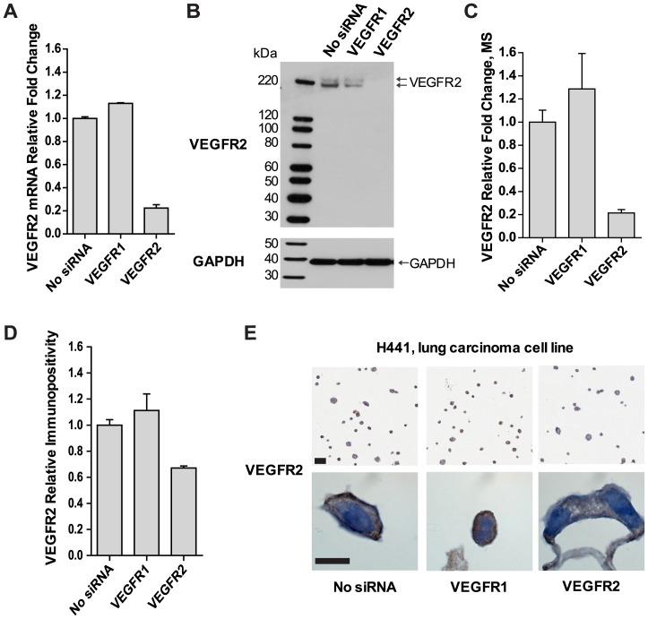
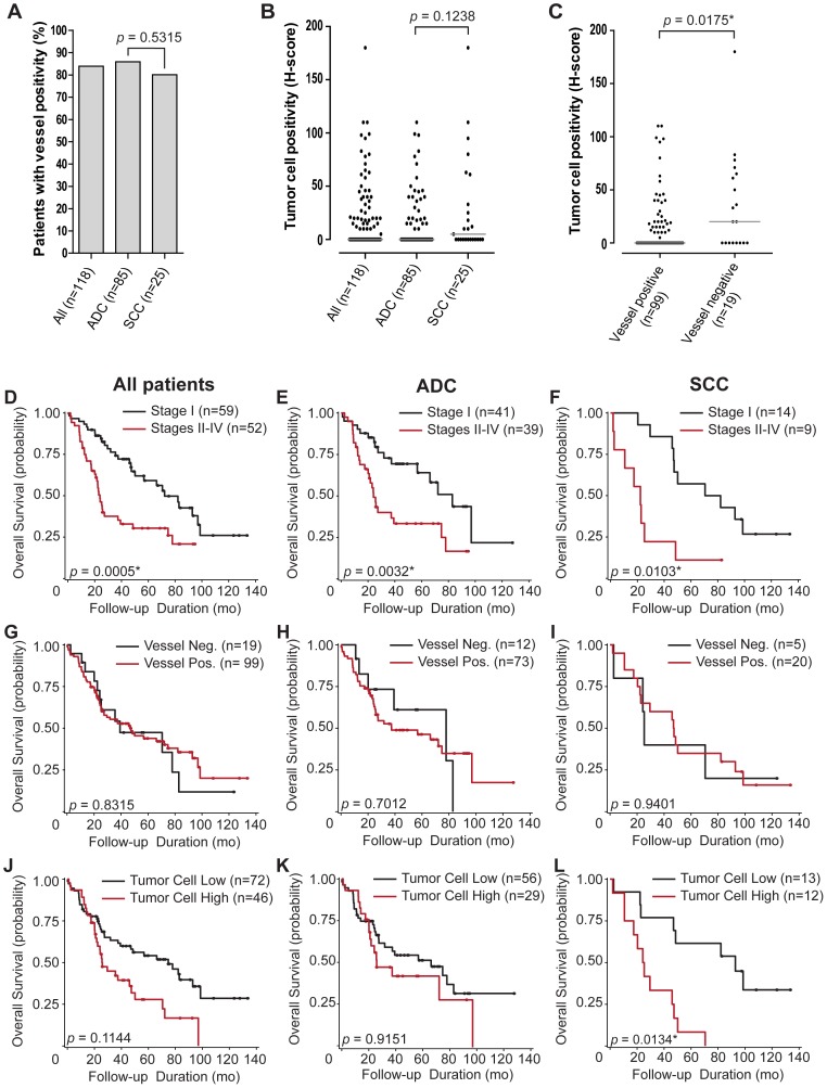
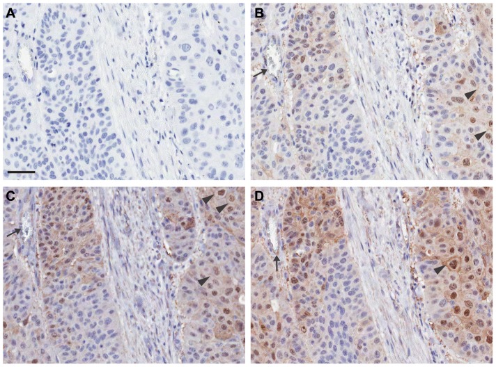
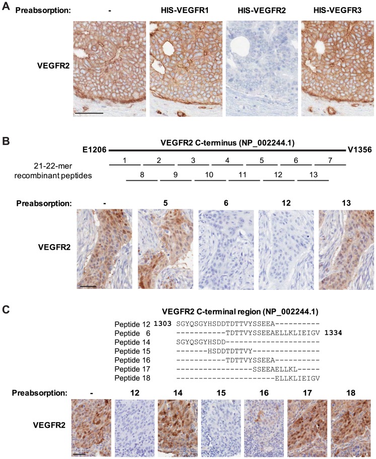
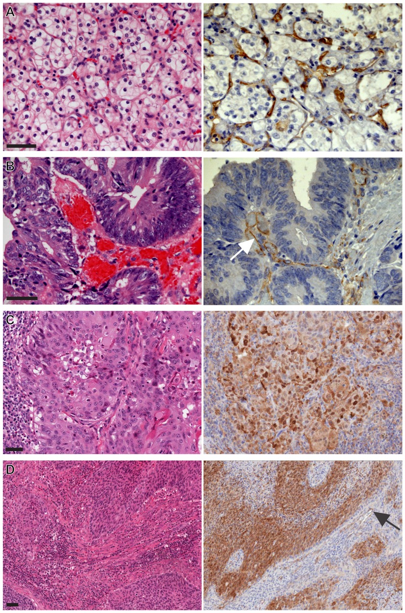
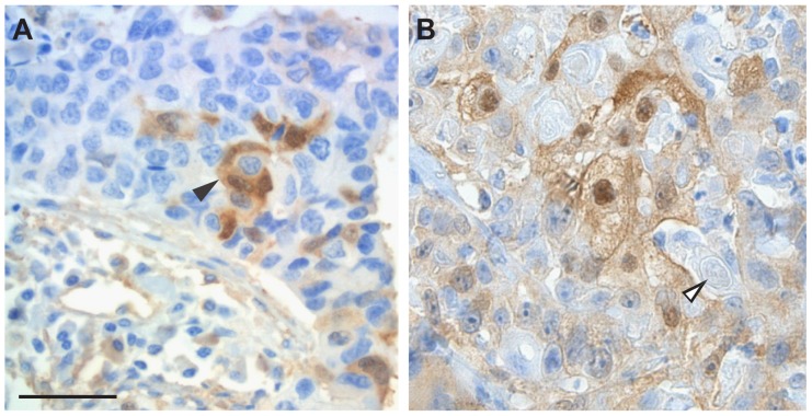
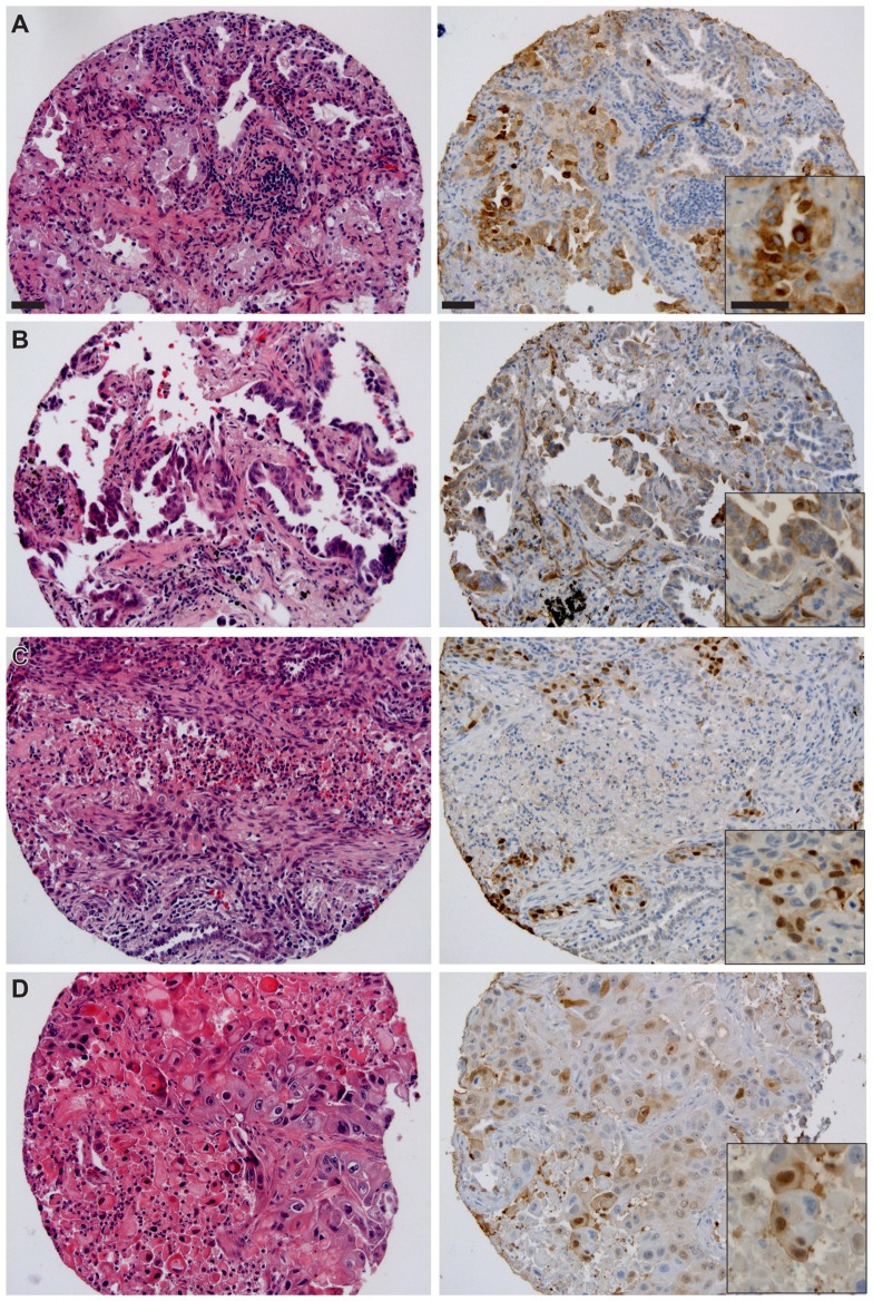
A robust immunohistochemical (IHC) assay for VEGFR2 was developed to investigate its utility for patient tailoring in clinical trials. The sensitivity, specificity, and selectivity of the IHC assay were established by siRNA knockdown, immunoblotting, mass spectrometry, and pre-absorption experiments. Characterization of the assay included screening a panel of multiple human cancer tissues and an independent cohort of non-small cell lung carcinoma (NSCLC, n = 118) characterized by TTF-1, p63, CK5/6, and CK7 IHC. VEGFR2 immunoreactivity was interpreted qualitatively (VEGFR2 positive/negative) in blood vessels and by semi-quantitative evaluation using H-scores in tumor cells (0-300). Associations were determined among combinations of VEGFR2 expression in blood vessels and tumor cells, and clinico-pathologic characteristics (age, sex, race, histologic subtype, disease stage) and overall survival using Kaplan-Meier analyses and appropriate statistical models. VEGFR2 expression both in blood vessels and in tumor cells in carcinomas of the lung, cervix, larynx, breast, and others was demonstrated. In the validation cohort, 99/118 (83.9%) NSCLC tissues expressed VEGFR2 in the blood vessels and 46/118 (39.0%) showed high tumor cell positivity (H-score ≥10). Vascular and tumor cell expression were inversely correlated (p = 0.0175). High tumor cell expression of VEGFR2 was associated with a 3.7-fold reduction in median overall survival in lung squamous-cell carcinoma (SCC, n = 25, p = 0.0134). The inverse correlation between vascular and tumor cell expression of VEGFR2 and the adverse prognosis associated with high VEGFR2 expression in immunohistochemically characterized pulmonary SCC are new findings with potential therapeutic implications. The robustness of this novel IHC assay will support further evaluation of its utility for patient tailoring in clinical trials of antiangiogenic agents.
开发了一种用于血管内皮生长因子受体2(VEGFR2)的强大免疫组织化学(IHC)检测方法,以研究其在临床试验中用于患者个体化治疗的效用。通过小干扰RNA(siRNA)敲低、免疫印迹、质谱分析和预吸附实验确定了该IHC检测方法的敏感性、特异性和选择性。该检测方法的特性包括筛选一组多种人类癌症组织以及一个以甲状腺转录因子-1(TTF-1)、p63、细胞角蛋白5/6(CK5/6)和细胞角蛋白7(CK7)免疫组织化学为特征的非小细胞肺癌(NSCLC,n = 118)独立队列。VEGFR2免疫反应性在血管中进行定性解释(VEGFR2阳性/阴性),并在肿瘤细胞中使用H评分进行半定量评估(0 - 300)。使用Kaplan-Meier分析和适当的统计模型确定血管和肿瘤细胞中VEGFR2表达组合与临床病理特征(年龄、性别、种族、组织学亚型、疾病分期)及总生存期之间的关联。在肺癌、宫颈癌、喉癌、乳腺癌及其他癌症中均证实了VEGFR2在血管和肿瘤细胞中的表达。在验证队列中,99/118(83.9%)的NSCLC组织在血管中表达VEGFR2,46/118(39.0%)显示肿瘤细胞高阳性(H评分≥10)。血管和肿瘤细胞表达呈负相关(p = 0.0175)。在肺鳞状细胞癌(SCC,n = 25)中,VEGFR2的高肿瘤细胞表达与中位总生存期降低3.7倍相关(p = 0.0134)。VEGFR2在血管和肿瘤细胞表达之间的负相关以及免疫组织化学特征化的肺SCC中高VEGFR2表达与不良预后相关是具有潜在治疗意义的新发现。这种新型IHC检测方法的稳健性将支持进一步评估其在抗血管生成药物临床试验中用于患者个体化治疗的效用。