1] Department of Molecular and Cellular Physiology, Stanford University School of Medicine, 279 Campus Drive, Stanford, California 94305, USA [2].
Nature. 2013 Dec 5;504(7478):101-6. doi: 10.1038/nature12735. Epub 2013 Nov 20.
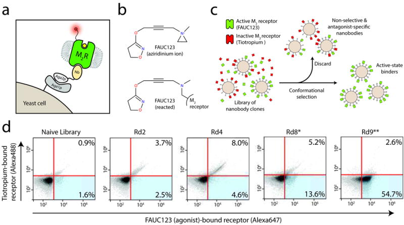
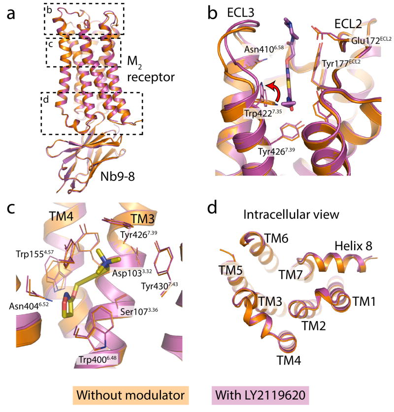
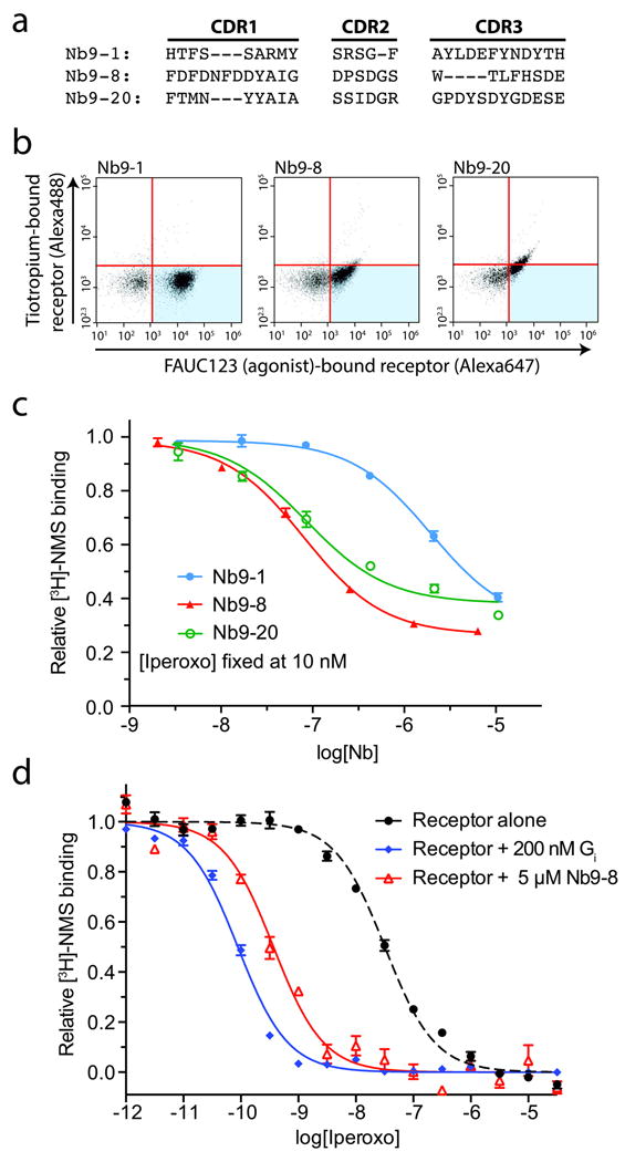
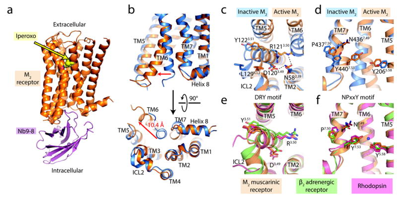
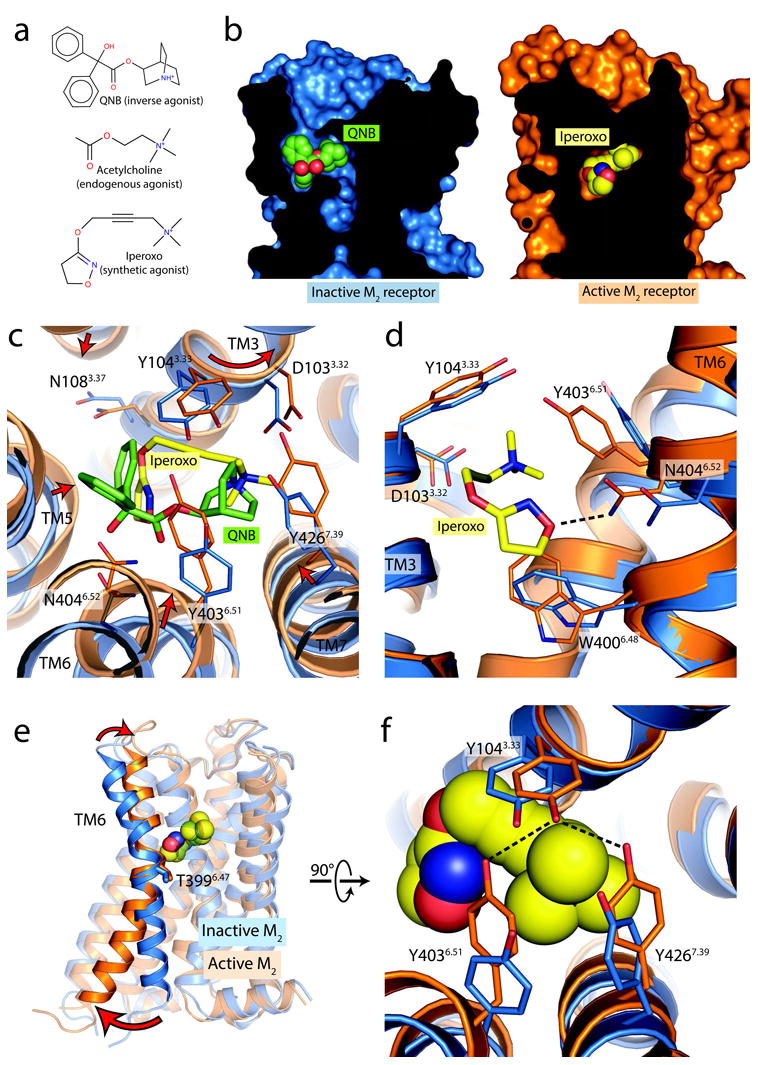
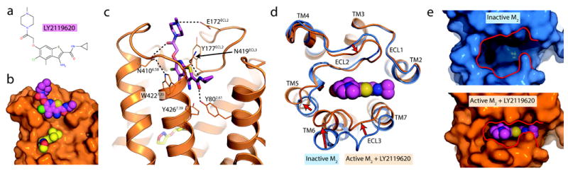
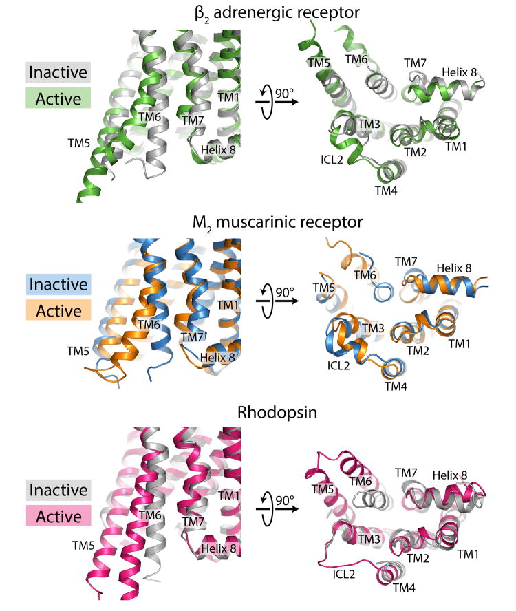
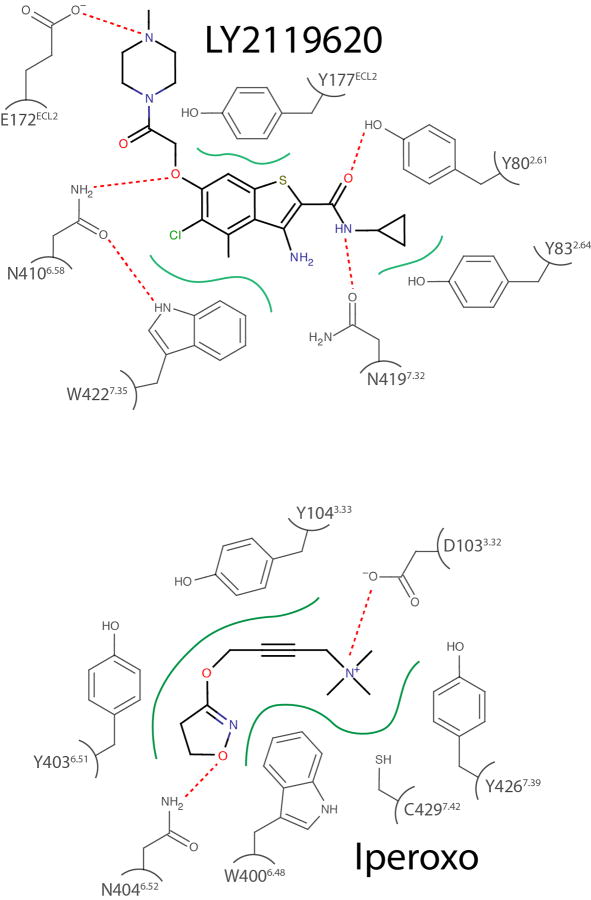
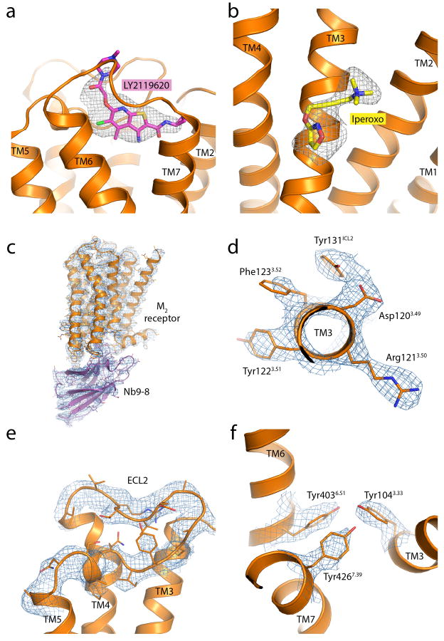
Despite recent advances in crystallography and the availability of G-protein-coupled receptor (GPCR) structures, little is known about the mechanism of their activation process, as only the β2 adrenergic receptor (β2AR) and rhodopsin have been crystallized in fully active conformations. Here we report the structure of an agonist-bound, active state of the human M2 muscarinic acetylcholine receptor stabilized by a G-protein mimetic camelid antibody fragment isolated by conformational selection using yeast surface display. In addition to the expected changes in the intracellular surface, the structure reveals larger conformational changes in the extracellular region and orthosteric binding site than observed in the active states of the β2AR and rhodopsin. We also report the structure of the M2 receptor simultaneously bound to the orthosteric agonist iperoxo and the positive allosteric modulator LY2119620. This structure reveals that LY2119620 recognizes a largely pre-formed binding site in the extracellular vestibule of the iperoxo-bound receptor, inducing a slight contraction of this outer binding pocket. These structures offer important insights into the activation mechanism and allosteric modulation of muscarinic receptors.
尽管结晶学最近取得了进展,并且可以获得 G 蛋白偶联受体 (GPCR) 的结构,但人们对它们的激活过程的机制知之甚少,因为只有β2 肾上腺素能受体 (β2AR) 和视紫红质以完全活跃的构象结晶。在这里,我们报告了一种激动剂结合的、由通过使用酵母表面展示进行构象选择分离的 G 蛋白模拟骆驼抗体片段稳定的人 M2 毒蕈碱乙酰胆碱受体的活性状态的结构。除了细胞内表面的预期变化外,该结构显示出比在β2AR 和视紫红质的活性状态中观察到的更大的细胞外区域和正构结合位点的构象变化。我们还报告了同时结合正构激动剂iperoxo 和正变构调节剂 LY2119620 的 M2 受体的结构。该结构表明,LY2119620 识别 iperoxo 结合受体细胞外前庭中预先形成的结合位点,诱导该外部结合口袋的轻微收缩。这些结构为毒蕈碱受体的激活机制和变构调节提供了重要的见解。