• 文献检索
  • 文档翻译
  • 深度研究
  • 学术资讯
  • Suppr Zotero 插件Zotero 插件
  • 邀请有礼
  • 套餐&价格
  • 历史记录
应用&插件
Suppr Zotero 插件Zotero 插件浏览器插件Mac 客户端Windows 客户端微信小程序
定价
高级版会员购买积分包购买API积分包
服务
文献检索文档翻译深度研究API 文档MCP 服务
关于我们
关于 Suppr公司介绍联系我们用户协议隐私条款
关注我们

Suppr 超能文献

核心技术专利:CN118964589B侵权必究
粤ICP备2023148730 号-1Suppr @ 2026

文献检索

告别复杂PubMed语法,用中文像聊天一样搜索,搜遍4000万医学文献。AI智能推荐,让科研检索更轻松。

立即免费搜索

文件翻译

保留排版,准确专业,支持PDF/Word/PPT等文件格式,支持 12+语言互译。

免费翻译文档

深度研究

AI帮你快速写综述,25分钟生成高质量综述,智能提取关键信息,辅助科研写作。

立即免费体验

基于体内人脑数据的扩散磁共振成像 compartment 模型排名。

A ranking of diffusion MRI compartment models with in vivo human brain data.

作者信息

Ferizi Uran, Schneider Torben, Panagiotaki Eleftheria, Nedjati-Gilani Gemma, Zhang Hui, Wheeler-Kingshott Claudia A M, Alexander Daniel C

机构信息

Department of Computer Science and Centre for Medical Image Computing, University College London, London, UK; NMR Research Unit, Department of Neuroinflammation, Institute of Neurology, University College London, London, UK.

出版信息

Magn Reson Med. 2014 Dec;72(6):1785-92. doi: 10.1002/mrm.25080. Epub 2013 Dec 17.

DOI:10.1002/mrm.25080
PMID:24347370
原文链接:https://pmc.ncbi.nlm.nih.gov/articles/PMC4278549/
Abstract

PURPOSE

Diffusion magnetic resonance imaging (MRI) microstructure imaging provides a unique noninvasive probe into tissue microstructure. The technique relies on biophysically motivated mathematical models, relating microscopic tissue features to the magnetic resonance (MR) signal. This work aims to determine which compartment models of diffusion MRI are best at describing measurements from in vivo human brain white matter.

METHODS

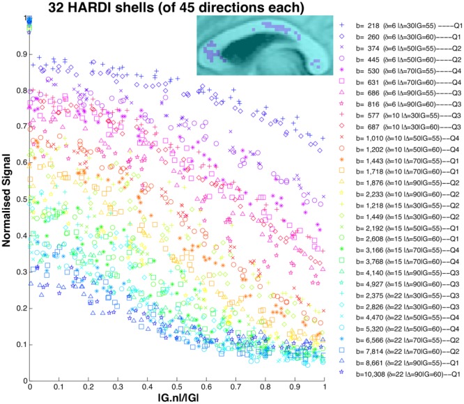
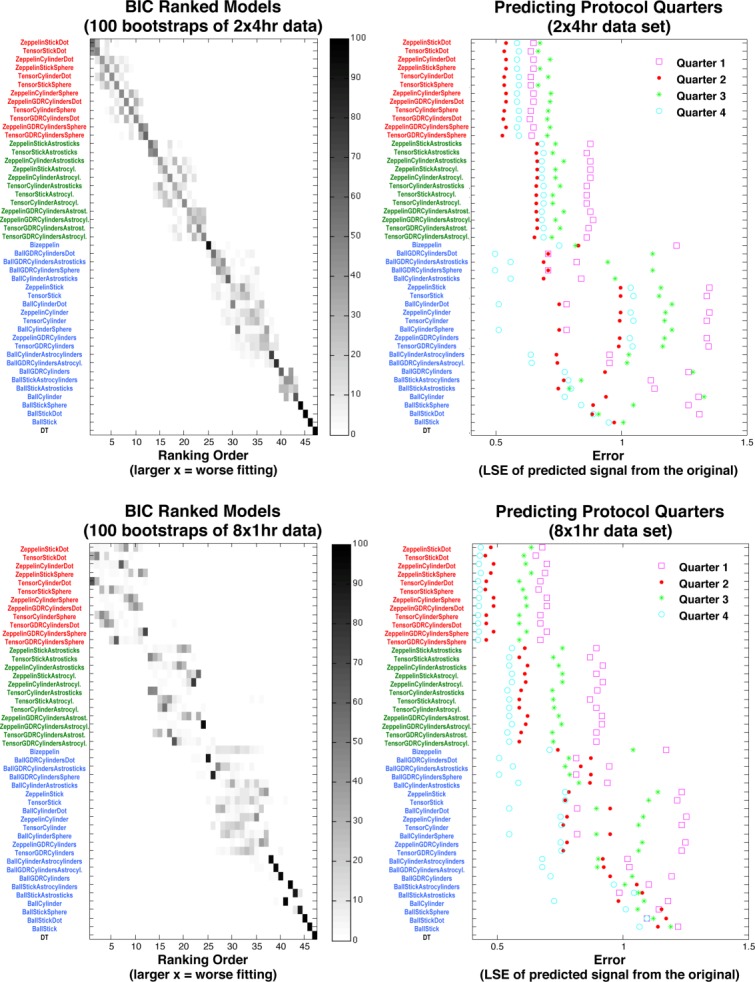
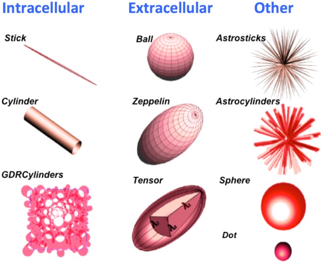
Recent work shows that three compartment models, designed to capture intra-axonal, extracellular, and isotropically restricted diffusion, best explain multi-b-value data sets from fixed rat corpus callosum. We extend this investigation to in vivo by using a live human subject on a clinical scanner. The analysis compares models of one, two, and three compartments and ranks their ability to explain the measured data. We enhance the original methodology to further evaluate the stability of the ranking.

RESULTS

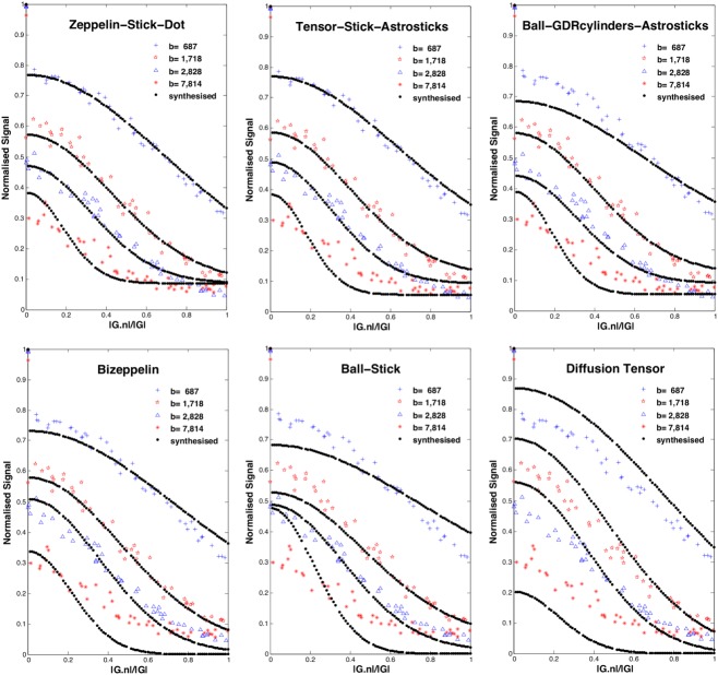
As with fixed tissue, three compartment models explain the data best. However, a clearer hierarchical structure and simpler models emerge. We also find that splitting the scanning into shorter sessions has little effect on the ranking of models, and that the results are broadly reproducible across sessions.

CONCLUSION

Three compartments are required to explain diffusion MR measurements from in vivo corpus callosum, which informs the choice of model for microstructure imaging applications in the brain.

摘要

目的

扩散磁共振成像(MRI)微观结构成像为研究组织微观结构提供了一种独特的非侵入性手段。该技术依赖于基于生物物理学原理的数学模型,将微观组织特征与磁共振(MR)信号联系起来。本研究旨在确定哪种扩散MRI的房室模型最能描述活体人类脑白质的测量结果。

方法

近期研究表明,旨在捕捉轴突内、细胞外和各向同性受限扩散的三室模型,能最好地解释来自固定大鼠胼胝体的多b值数据集。我们通过在临床扫描仪上对一名活体人类受试者进行研究,将这一调查扩展到活体研究。分析比较了一室、二室和三室模型,并对它们解释测量数据的能力进行排名。我们改进了原始方法,以进一步评估排名的稳定性。

结果

与固定组织一样,三室模型对数据的解释效果最佳。然而,出现了更清晰的层次结构和更简单的模型。我们还发现,将扫描分成较短的时间段对模型排名影响不大,并且结果在各时间段之间具有广泛的可重复性。

结论

需要三室模型来解释活体胼胝体的扩散MR测量结果,这为脑部微观结构成像应用中的模型选择提供了依据。

https://cdn.ncbi.nlm.nih.gov/pmc/blobs/0fb0/4278549/5be45abe8526/mrm0072-1785-f4.jpg
https://cdn.ncbi.nlm.nih.gov/pmc/blobs/0fb0/4278549/090e0db1c625/mrm0072-1785-f1.jpg
https://cdn.ncbi.nlm.nih.gov/pmc/blobs/0fb0/4278549/beedfd214f68/mrm0072-1785-f2.jpg
https://cdn.ncbi.nlm.nih.gov/pmc/blobs/0fb0/4278549/c03bcd5a1ba5/mrm0072-1785-f3.jpg
https://cdn.ncbi.nlm.nih.gov/pmc/blobs/0fb0/4278549/5be45abe8526/mrm0072-1785-f4.jpg

相似文献

1
A ranking of diffusion MRI compartment models with in vivo human brain data.基于体内人脑数据的扩散磁共振成像 compartment 模型排名。
Magn Reson Med. 2014 Dec;72(6):1785-92. doi: 10.1002/mrm.25080. Epub 2013 Dec 17.
2
Two-compartment models of the diffusion MR signal in brain white matter.脑白质中扩散磁共振信号的双室模型。
Med Image Comput Comput Assist Interv. 2009;12(Pt 1):329-36. doi: 10.1007/978-3-642-04268-3_41.
3
New perspectives on the sources of white matter DTI signal.白质扩散张量成像(DTI)信号来源的新视角。
IEEE Trans Med Imaging. 2007 Nov;26(11):1448-55. doi: 10.1109/TMI.2007.906787.
4
White matter compartment models for in vivo diffusion MRI at 300mT/m.用于300mT/m下体内扩散磁共振成像的白质区室模型。
Neuroimage. 2015 Sep;118:468-83. doi: 10.1016/j.neuroimage.2015.06.027. Epub 2015 Jun 16.
5
Understanding diffusion MR imaging techniques: from scalar diffusion-weighted imaging to diffusion tensor imaging and beyond.理解扩散磁共振成像技术:从标量扩散加权成像到扩散张量成像及其他。
Radiographics. 2006 Oct;26 Suppl 1:S205-23. doi: 10.1148/rg.26si065510.
6
Age-related alterations in axonal microstructure in the corpus callosum measured by high-gradient diffusion MRI.高梯度扩散 MRI 测量胼胝体轴突微观结构的年龄相关性改变。
Neuroimage. 2019 May 1;191:325-336. doi: 10.1016/j.neuroimage.2019.02.036. Epub 2019 Feb 18.
7
Assessing white matter microstructure of the newborn with multi-shell diffusion MRI and biophysical compartment models.应用多壳层扩散 MRI 和生物物理房室模型评估新生儿的脑白质微观结构。
Neuroimage. 2014 Aug 1;96:288-99. doi: 10.1016/j.neuroimage.2014.03.057. Epub 2014 Mar 28.
8
Characterizing brain tissue by assessment of the distribution of anisotropic microstructural environments in diffusion-compartment imaging (DIAMOND).通过评估扩散室成像(DIAMOND)中各向异性微观结构环境的分布来表征脑组织。
Magn Reson Med. 2016 Sep;76(3):963-77. doi: 10.1002/mrm.25912. Epub 2015 Sep 12.
9
Diffusion MRI microstructure models with in vivo human brain Connectome data: results from a multi-group comparison.具有体内人脑连接组数据的扩散磁共振成像微观结构模型:多组比较结果
NMR Biomed. 2017 Sep;30(9). doi: 10.1002/nbm.3734. Epub 2017 Jun 23.
10
Estimation of extracellular volume from regularized multi-shell diffusion MRI.基于正则化多壳扩散磁共振成像的细胞外体积估计
Med Image Comput Comput Assist Interv. 2012;15(Pt 2):305-12. doi: 10.1007/978-3-642-33418-4_38.

引用本文的文献

1
Multi-tensor fixel-based metrics in tractometry: application to multiple sclerosis.基于多张量体素的纤维束测量指标:在多发性硬化症中的应用
Front Neurosci. 2024 Dec 20;18:1467786. doi: 10.3389/fnins.2024.1467786. eCollection 2024.
2
MR Imaging in Ataxias: Consensus Recommendations by the Ataxia Global Initiative Working Group on MRI Biomarkers.磁共振成像在共济失调中的应用:共济失调全球倡议磁共振成像生物标志物工作组的共识建议。
Cerebellum. 2024 Jun;23(3):931-945. doi: 10.1007/s12311-023-01572-y. Epub 2023 Jun 6.
3
Linear multi-scale modeling of diffusion MRI data: A framework for characterization of oriented structures across length scales.

本文引用的文献

1
Contrast and stability of the axon diameter index from microstructure imaging with diffusion MRI.基于扩散磁共振成像的微观结构成像中轴突直径指数的对比度与稳定性
Magn Reson Med. 2013 Sep;70(3):711-21. doi: 10.1002/mrm.24501. Epub 2012 Sep 28.
2
NODDI: practical in vivo neurite orientation dispersion and density imaging of the human brain.NODDI:人脑神经丝取向分散和密度成像的实用体内技术。
Neuroimage. 2012 Jul 16;61(4):1000-16. doi: 10.1016/j.neuroimage.2012.03.072. Epub 2012 Mar 30.
3
Ball and rackets: Inferring fiber fanning from diffusion-weighted MRI.
线性多尺度扩散磁共振成像数据建模:一种跨尺度描述各向异性结构特征的方法。
Hum Brain Mapp. 2023 Mar;44(4):1496-1514. doi: 10.1002/hbm.26143. Epub 2022 Dec 7.
4
Accuracy and reliability of diffusion imaging models.弥散成像模型的准确性和可靠性。
Neuroimage. 2022 Jul 1;254:119138. doi: 10.1016/j.neuroimage.2022.119138. Epub 2022 Mar 23.
5
Constrained spherical deconvolution of nonspherically sampled diffusion MRI data.约束球面反卷积非球采样扩散 MRI 数据。
Hum Brain Mapp. 2021 Feb 1;42(2):521-538. doi: 10.1002/hbm.25241. Epub 2020 Nov 10.
6
Towards unconstrained compartment modeling in white matter using diffusion-relaxation MRI with tensor-valued diffusion encoding.利用张量值扩散编码的扩散弛豫磁共振成像实现白质中无约束的隔室建模。
Magn Reson Med. 2020 Sep;84(3):1605-1623. doi: 10.1002/mrm.28216. Epub 2020 Mar 6.
7
The dot-compartment revealed? Diffusion MRI with ultra-strong gradients and spherical tensor encoding in the living human brain.点分辨?活体人脑中超强梯度和球形张量编码的扩散 MRI。
Neuroimage. 2020 Apr 15;210:116534. doi: 10.1016/j.neuroimage.2020.116534. Epub 2020 Jan 11.
8
Comparison of q-Space Reconstruction Methods for Undersampled Diffusion Spectrum Imaging Data.基于欠采样的扩散谱成像数据的 q-Space 重建方法比较。
Magn Reson Med Sci. 2020 May 1;19(2):108-118. doi: 10.2463/mrms.mp.2019-0015. Epub 2019 May 10.
9
Microscopic anisotropy misestimation in spherical-mean single diffusion encoding MRI.球谐平均单扩散编码 MRI 中微观各向异性的估计误差。
Magn Reson Med. 2019 May;81(5):3245-3261. doi: 10.1002/mrm.27606. Epub 2019 Jan 16.
10
Review of diffusion MRI studies in chronic white matter diseases.慢性白质疾病的扩散磁共振成像研究综述
Neurosci Lett. 2019 Feb 16;694:198-207. doi: 10.1016/j.neulet.2018.12.007. Epub 2018 Dec 6.
球和球拍:从弥散加权 MRI 推断纤维扇面。
Neuroimage. 2012 Apr 2;60(2):1412-25. doi: 10.1016/j.neuroimage.2012.01.056. Epub 2012 Jan 14.
4
The importance of axonal undulation in diffusion MR measurements: a Monte Carlo simulation study.轴突波动在扩散磁共振测量中的重要性:一项蒙特卡罗模拟研究。
NMR Biomed. 2012 May;25(5):795-805. doi: 10.1002/nbm.1795. Epub 2011 Oct 21.
5
Compartment models of the diffusion MR signal in brain white matter: a taxonomy and comparison.脑白质磁共振扩散信号的隔室模型:分类与比较。
Neuroimage. 2012 Feb 1;59(3):2241-54. doi: 10.1016/j.neuroimage.2011.09.081. Epub 2011 Oct 7.
6
Axon diameter mapping in the presence of orientation dispersion with diffusion MRI.弥散磁共振成像中存在方向分散时的轴突直径测绘。
Neuroimage. 2011 Jun 1;56(3):1301-15. doi: 10.1016/j.neuroimage.2011.01.084. Epub 2011 Feb 19.
7
Orientationally invariant indices of axon diameter and density from diffusion MRI.基于弥散磁共振成像的各向同性轴突直径和密度指标
Neuroimage. 2010 Oct 1;52(4):1374-89. doi: 10.1016/j.neuroimage.2010.05.043. Epub 2010 May 23.
8
In vivo measurement of axon diameter distribution in the corpus callosum of rat brain.大鼠脑胼胝体轴突直径分布的体内测量
Brain. 2009 May;132(Pt 5):1210-20. doi: 10.1093/brain/awp042. Epub 2009 Apr 29.
9
Aldehyde fixative solutions alter the water relaxation and diffusion properties of nervous tissue.醛类固定剂溶液会改变神经组织的水弛豫和扩散特性。
Magn Reson Med. 2009 Jul;62(1):26-34. doi: 10.1002/mrm.21977.
10
A general framework for experiment design in diffusion MRI and its application in measuring direct tissue-microstructure features.扩散磁共振成像实验设计的通用框架及其在测量直接组织微观结构特征中的应用。
Magn Reson Med. 2008 Aug;60(2):439-48. doi: 10.1002/mrm.21646.