• 文献检索
  • 文档翻译
  • 深度研究
  • 学术资讯
  • Suppr Zotero 插件Zotero 插件
  • 邀请有礼
  • 套餐&价格
  • 历史记录
应用&插件
Suppr Zotero 插件Zotero 插件浏览器插件Mac 客户端Windows 客户端微信小程序
定价
高级版会员购买积分包购买API积分包
服务
文献检索文档翻译深度研究API 文档MCP 服务
关于我们
关于 Suppr公司介绍联系我们用户协议隐私条款
关注我们

Suppr 超能文献

核心技术专利:CN118964589B侵权必究
粤ICP备2023148730 号-1Suppr @ 2026

文献检索

告别复杂PubMed语法,用中文像聊天一样搜索,搜遍4000万医学文献。AI智能推荐,让科研检索更轻松。

立即免费搜索

文件翻译

保留排版,准确专业,支持PDF/Word/PPT等文件格式,支持 12+语言互译。

免费翻译文档

深度研究

AI帮你快速写综述,25分钟生成高质量综述,智能提取关键信息,辅助科研写作。

立即免费体验

具有体内人脑连接组数据的扩散磁共振成像微观结构模型:多组比较结果

Diffusion MRI microstructure models with in vivo human brain Connectome data: results from a multi-group comparison.

作者信息

Ferizi Uran, Scherrer Benoit, Schneider Torben, Alipoor Mohammad, Eufracio Odin, Fick Rutger H J, Deriche Rachid, Nilsson Markus, Loya-Olivas Ana K, Rivera Mariano, Poot Dirk H J, Ramirez-Manzanares Alonso, Marroquin Jose L, Rokem Ariel, Pötter Christian, Dougherty Robert F, Sakaie Ken, Wheeler-Kingshott Claudia, Warfield Simon K, Witzel Thomas, Wald Lawrence L, Raya José G, Alexander Daniel C

机构信息

Centre for Medical Image Computing, Department of Computer Science, University College London, UK.

Department of Radiology, New York University School of Medicine, USA.

出版信息

NMR Biomed. 2017 Sep;30(9). doi: 10.1002/nbm.3734. Epub 2017 Jun 23.

DOI:10.1002/nbm.3734
PMID:28643354
原文链接:https://pmc.ncbi.nlm.nih.gov/articles/PMC5563694/
Abstract

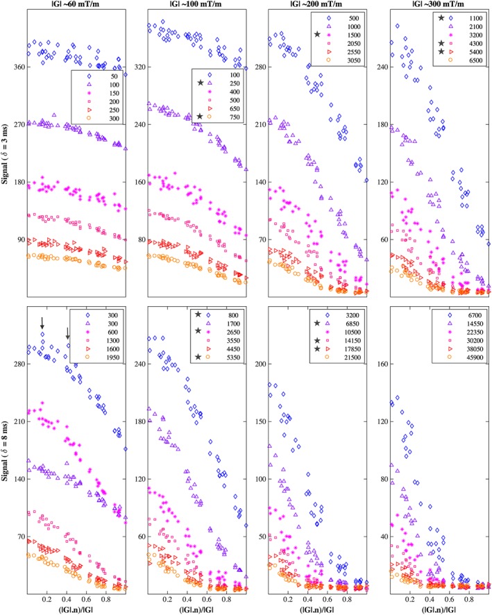
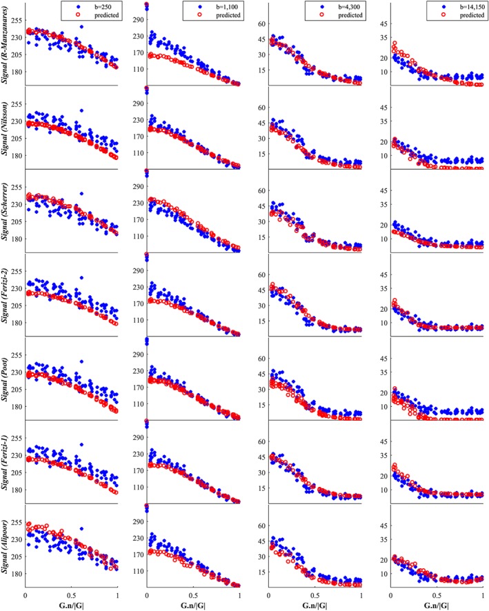
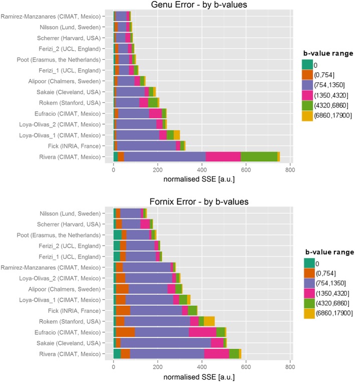
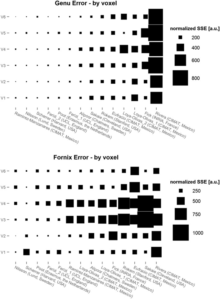
A large number of mathematical models have been proposed to describe the measured signal in diffusion-weighted (DW) magnetic resonance imaging (MRI). However, model comparison to date focuses only on specific subclasses, e.g. compartment models or signal models, and little or no information is available in the literature on how performance varies among the different types of models. To address this deficiency, we organized the 'White Matter Modeling Challenge' during the International Symposium on Biomedical Imaging (ISBI) 2015 conference. This competition aimed to compare a range of different kinds of models in their ability to explain a large range of measurable in vivo DW human brain data. Specifically, we assessed the ability of models to predict the DW signal accurately for new diffusion gradients and b values. We did not evaluate the accuracy of estimated model parameters, as a ground truth is hard to obtain. We used the Connectome scanner at the Massachusetts General Hospital, using gradient strengths of up to 300 mT/m and a broad set of diffusion times. We focused on assessing the DW signal prediction in two regions: the genu in the corpus callosum, where the fibres are relatively straight and parallel, and the fornix, where the configuration of fibres is more complex. The challenge participants had access to three-quarters of the dataset and their models were ranked on their ability to predict the remaining unseen quarter of the data. The challenge provided a unique opportunity for a quantitative comparison of diverse methods from multiple groups worldwide. The comparison of the challenge entries reveals interesting trends that could potentially influence the next generation of diffusion-based quantitative MRI techniques. The first is that signal models do not necessarily outperform tissue models; in fact, of those tested, tissue models rank highest on average. The second is that assuming a non-Gaussian (rather than purely Gaussian) noise model provides little improvement in prediction of unseen data, although it is possible that this may still have a beneficial effect on estimated parameter values. The third is that preprocessing the training data, here by omitting signal outliers, and using signal-predicting strategies, such as bootstrapping or cross-validation, could benefit the model fitting. The analysis in this study provides a benchmark for other models and the data remain available to build up a more complete comparison in the future.

摘要

已经提出了大量数学模型来描述扩散加权磁共振成像(DW-MRI)中的测量信号。然而,迄今为止的模型比较仅集中在特定的子类上,例如隔室模型或信号模型,并且文献中几乎没有关于不同类型模型之间性能如何变化的信息。为了弥补这一不足,我们在2015年国际生物医学成像研讨会(ISBI)会议期间组织了“白质建模挑战赛”。该竞赛旨在比较一系列不同类型的模型解释大量可测量的体内DW人脑数据的能力。具体来说,我们评估了模型针对新的扩散梯度和b值准确预测DW信号的能力。由于难以获得真实值,我们没有评估估计模型参数的准确性。我们使用了马萨诸塞州总医院的Connectome扫描仪,其梯度强度高达300 mT/m,并采用了广泛的扩散时间。我们专注于评估两个区域的DW信号预测:胼胝体的膝部,此处纤维相对笔直且平行;以及穹窿,此处纤维的构型更为复杂。挑战赛参与者可以使用四分之三的数据集,并根据他们预测其余四分之一未见数据的能力对其模型进行排名。该挑战赛为全球多个团队的不同方法进行定量比较提供了独特的机会。对挑战赛参赛作品的比较揭示了一些有趣的趋势,这些趋势可能会对下一代基于扩散的定量MRI技术产生潜在影响。第一个趋势是信号模型不一定优于组织模型;事实上,在测试的模型中,组织模型的平均排名最高。第二个趋势是,假设非高斯(而不是纯高斯)噪声模型在预测未见数据方面几乎没有改进,尽管这仍有可能对估计的参数值产生有益影响。第三个趋势是,对训练数据进行预处理,在这里是通过省略信号异常值,并使用诸如自助法或交叉验证等信号预测策略,可能会有利于模型拟合。本研究中的分析为其他模型提供了一个基准,并且这些数据仍然可用,以便在未来进行更全面的比较。

https://cdn.ncbi.nlm.nih.gov/pmc/blobs/22f0/5575565/05328ac4adc7/NBM-30-na-g008.jpg
https://cdn.ncbi.nlm.nih.gov/pmc/blobs/22f0/5575565/be4255e8740b/NBM-30-na-g002.jpg
https://cdn.ncbi.nlm.nih.gov/pmc/blobs/22f0/5575565/3b1ee5318a39/NBM-30-na-g004.jpg
https://cdn.ncbi.nlm.nih.gov/pmc/blobs/22f0/5575565/bf01a8af2c62/NBM-30-na-g005.jpg
https://cdn.ncbi.nlm.nih.gov/pmc/blobs/22f0/5575565/05328ac4adc7/NBM-30-na-g008.jpg

相似文献

1
Diffusion MRI microstructure models with in vivo human brain Connectome data: results from a multi-group comparison.具有体内人脑连接组数据的扩散磁共振成像微观结构模型:多组比较结果
NMR Biomed. 2017 Sep;30(9). doi: 10.1002/nbm.3734. Epub 2017 Jun 23.
2
Age-related alterations in axonal microstructure in the corpus callosum measured by high-gradient diffusion MRI.高梯度扩散 MRI 测量胼胝体轴突微观结构的年龄相关性改变。
Neuroimage. 2019 May 1;191:325-336. doi: 10.1016/j.neuroimage.2019.02.036. Epub 2019 Feb 18.
3
Connectome 2.0: Developing the next-generation ultra-high gradient strength human MRI scanner for bridging studies of the micro-, meso- and macro-connectome.连接组学 2.0:开发下一代超高梯度场强的人类 MRI 扫描仪,以连接微观、介观和宏观连接组学的研究。
Neuroimage. 2021 Nov;243:118530. doi: 10.1016/j.neuroimage.2021.118530. Epub 2021 Aug 28.
4
White matter compartment models for in vivo diffusion MRI at 300mT/m.用于300mT/m下体内扩散磁共振成像的白质区室模型。
Neuroimage. 2015 Sep;118:468-83. doi: 10.1016/j.neuroimage.2015.06.027. Epub 2015 Jun 16.
5
Linear multi-scale modeling of diffusion MRI data: A framework for characterization of oriented structures across length scales.线性多尺度扩散磁共振成像数据建模:一种跨尺度描述各向异性结构特征的方法。
Hum Brain Mapp. 2023 Mar;44(4):1496-1514. doi: 10.1002/hbm.26143. Epub 2022 Dec 7.
6
Mapping the human connectome using diffusion MRI at 300 mT/m gradient strength: Methodological advances and scientific impact.使用 300 mT/m 梯度强度的弥散 MRI 绘制人类连接组图谱:方法学进展和科学影响。
Neuroimage. 2022 Jul 1;254:118958. doi: 10.1016/j.neuroimage.2022.118958. Epub 2022 Feb 23.
7
In vivo cortical microstructure mapping using high-gradient diffusion MRI accounting for intercompartmental water exchange effects.利用考虑隔室间水交换效应的高梯度扩散磁共振成像进行体内皮质微结构映射。
Neuroimage. 2025 Jul 1;314:121258. doi: 10.1016/j.neuroimage.2025.121258. Epub 2025 May 9.
8
Fiber estimation and tractography in diffusion MRI: development of simulated brain images and comparison of multi-fiber analysis methods at clinical b-values.扩散磁共振成像中的纤维估计与纤维束成像:模拟脑图像的开发及临床b值下多纤维分析方法的比较
Neuroimage. 2015 Apr 1;109:341-56. doi: 10.1016/j.neuroimage.2014.12.060. Epub 2014 Dec 30.
9
Generalized Richardson-Lucy (GRL) for analyzing multi-shell diffusion MRI data.用于分析多壳扩散磁共振成像数据的广义理查森-露西(GRL)算法
Neuroimage. 2020 Sep;218:116948. doi: 10.1016/j.neuroimage.2020.116948. Epub 2020 May 16.
10
A ranking of diffusion MRI compartment models with in vivo human brain data.基于体内人脑数据的扩散磁共振成像 compartment 模型排名。
Magn Reson Med. 2014 Dec;72(6):1785-92. doi: 10.1002/mrm.25080. Epub 2013 Dec 17.

引用本文的文献

1
Ultra-high gradient connectomics and microstructure MRI scanner for imaging of human brain circuits across scales.用于跨尺度成像人类脑回路的超高梯度连接组学和微观结构MRI扫描仪。
Nat Biomed Eng. 2025 Jul 16. doi: 10.1038/s41551-025-01457-x.
2
What has brain diffusion magnetic resonance imaging taught us about chronic primary pain: a narrative review.脑扩散磁共振成像对慢性原发性疼痛有何启示:一项叙述性综述
Pain. 2025 Feb 1;166(2):243-261. doi: 10.1097/j.pain.0000000000003345. Epub 2024 Aug 21.
3
Multi-tensor fixel-based metrics in tractometry: application to multiple sclerosis.

本文引用的文献

1
Multi-compartment microscopic diffusion imaging.多室微观扩散成像
Neuroimage. 2016 Oct 1;139:346-359. doi: 10.1016/j.neuroimage.2016.06.002. Epub 2016 Jun 6.
2
In vivo observation and biophysical interpretation of time-dependent diffusion in human white matter.人体白质中时间依赖性扩散的体内观察与生物物理解释
Neuroimage. 2016 Apr 1;129:414-427. doi: 10.1016/j.neuroimage.2016.01.018. Epub 2016 Jan 20.
3
MRBrainS Challenge: Online Evaluation Framework for Brain Image Segmentation in 3T MRI Scans.MRBrainS挑战赛:3T磁共振成像扫描中脑图像分割的在线评估框架
基于多张量体素的纤维束测量指标:在多发性硬化症中的应用
Front Neurosci. 2024 Dec 20;18:1467786. doi: 10.3389/fnins.2024.1467786. eCollection 2024.
4
Resolution and b value dependent structural connectome in ex vivo mouse brain.离体小鼠脑的分辨率和 b 值相关结构连接组学
Neuroimage. 2022 Jul 15;255:119199. doi: 10.1016/j.neuroimage.2022.119199. Epub 2022 Apr 10.
5
Mapping the human connectome using diffusion MRI at 300 mT/m gradient strength: Methodological advances and scientific impact.使用 300 mT/m 梯度强度的弥散 MRI 绘制人类连接组图谱:方法学进展和科学影响。
Neuroimage. 2022 Jul 1;254:118958. doi: 10.1016/j.neuroimage.2022.118958. Epub 2022 Feb 23.
6
Development of white matter microstructure and executive functions during childhood and adolescence: a review of diffusion MRI studies.儿童和青少年时期脑白质微观结构和执行功能的发展:弥散磁共振成像研究综述。
Dev Cogn Neurosci. 2021 Oct;51:101008. doi: 10.1016/j.dcn.2021.101008. Epub 2021 Aug 25.
7
Age-Related Variations in Regional White Matter Volumetry and Microstructure During the Post-adolescence Period: A Cross-Sectional Study of a Cohort of 1,713 University Students.青春期后阶段区域白质体积测定和微观结构的年龄相关变化:一项对1713名大学生队列的横断面研究。
Front Syst Neurosci. 2021 Aug 3;15:692152. doi: 10.3389/fnsys.2021.692152. eCollection 2021.
8
On the generalizability of diffusion MRI signal representations across acquisition parameters, sequences and tissue types: Chronicles of the MEMENTO challenge.弥散磁共振成像信号表示在采集参数、序列和组织类型上的可推广性:MEMENTO 挑战赛纪事。
Neuroimage. 2021 Oct 15;240:118367. doi: 10.1016/j.neuroimage.2021.118367. Epub 2021 Jul 6.
9
Alcohol consumption in the general population is associated with structural changes in multiple organ systems.普通人群的饮酒与多个器官系统的结构变化有关。
Elife. 2021 Jun 1;10:e65325. doi: 10.7554/eLife.65325.
10
Quantitative magnetic resonance imaging towards clinical application in multiple sclerosis.定量磁共振成像在多发性硬化症中的临床应用。
Brain. 2021 Jun 22;144(5):1296-1311. doi: 10.1093/brain/awab029.
Comput Intell Neurosci. 2015;2015:813696. doi: 10.1155/2015/813696. Epub 2015 Dec 2.
4
Degeneracy in model parameter estimation for multi-compartmental diffusion in neuronal tissue.神经元组织中多房室扩散模型参数估计的简并性。
NMR Biomed. 2016 Jan;29(1):33-47. doi: 10.1002/nbm.3450. Epub 2015 Nov 29.
5
Sparse Reconstruction Challenge for diffusion MRI: Validation on a physical phantom to determine which acquisition scheme and analysis method to use?扩散磁共振成像的稀疏重建挑战:在物理体模上进行验证以确定使用哪种采集方案和分析方法?
Med Image Anal. 2015 Dec;26(1):316-31. doi: 10.1016/j.media.2015.10.012. Epub 2015 Nov 10.
6
Diffusion kurtosis imaging probes cortical alterations and white matter pathology following cuprizone induced demyelination and spontaneous remyelination.扩散峰度成像探究了铜离子螯合剂诱导脱髓鞘和自发髓鞘再生后的皮质改变和白质病变。
Neuroimage. 2016 Jan 15;125:363-377. doi: 10.1016/j.neuroimage.2015.10.052. Epub 2015 Oct 23.
7
Sparse and Adaptive Diffusion Dictionary (SADD) for recovering intra-voxel white matter structure.用于恢复体素内白质结构的稀疏和自适应扩散字典 (SADD)。
Med Image Anal. 2015 Dec;26(1):243-55. doi: 10.1016/j.media.2015.10.002. Epub 2015 Oct 22.
8
Evaluation of diffusion kurtosis imaging in ex vivo hypomyelinated mouse brains.脱髓鞘小鼠离体脑扩散峰度成像评估
Neuroimage. 2016 Jan 1;124(Pt A):612-626. doi: 10.1016/j.neuroimage.2015.09.028. Epub 2015 Sep 21.
9
Characterizing brain tissue by assessment of the distribution of anisotropic microstructural environments in diffusion-compartment imaging (DIAMOND).通过评估扩散室成像(DIAMOND)中各向异性微观结构环境的分布来表征脑组织。
Magn Reson Med. 2016 Sep;76(3):963-77. doi: 10.1002/mrm.25912. Epub 2015 Sep 12.
10
The DTI Challenge: Toward Standardized Evaluation of Diffusion Tensor Imaging Tractography for Neurosurgery.DTI 挑战:迈向神经外科手术中扩散张量成像纤维束成像的标准化评估
J Neuroimaging. 2015 Nov-Dec;25(6):875-82. doi: 10.1111/jon.12283. Epub 2015 Aug 11.