Thayer School of Engineering at Dartmouth, Hanover, New Hampshire, United States of America.
PLoS One. 2013 Dec 23;8(12):e83299. doi: 10.1371/journal.pone.0083299. eCollection 2013.
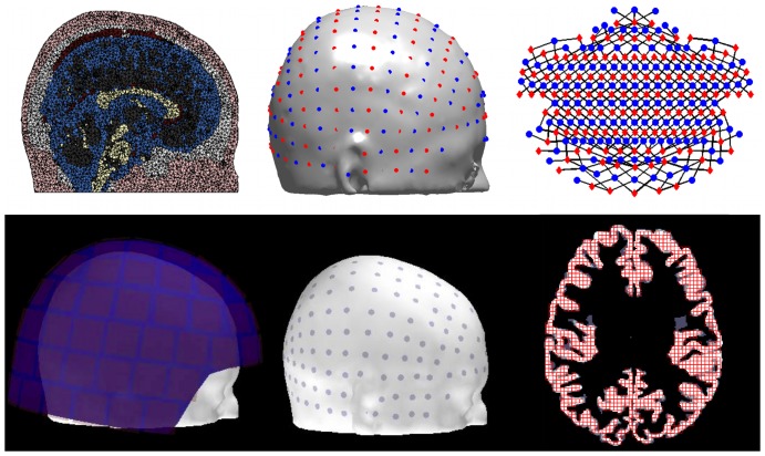
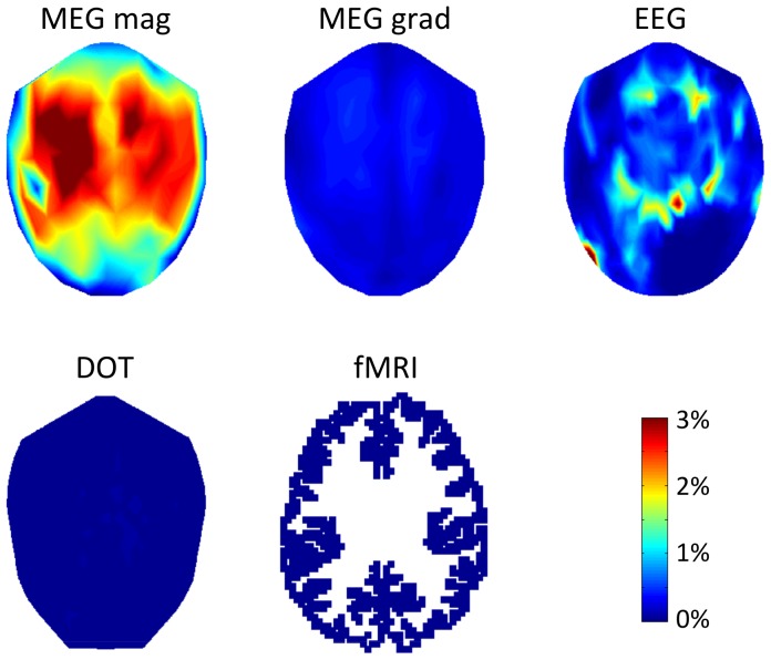
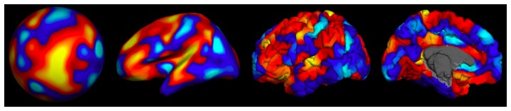
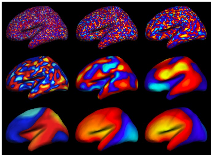
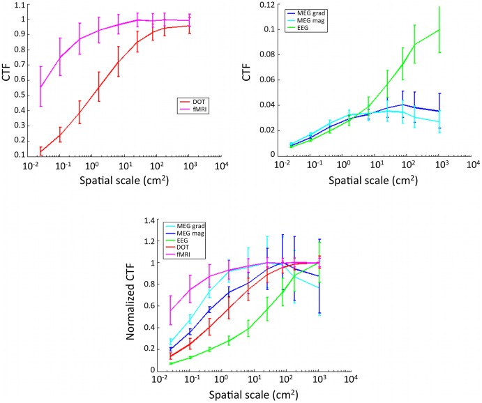
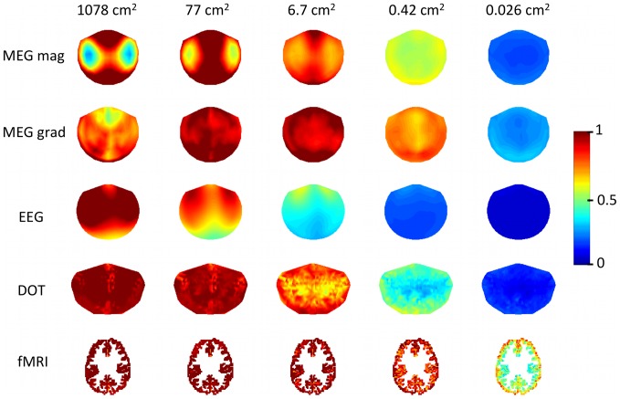
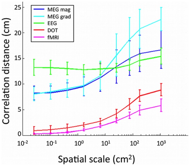
The objective of this work is to quantify how patterns of cortical activity at different spatial scales are measured by noninvasive functional neuroimaging sensors. We simulated cortical activation patterns at nine different spatial scales in a realistic head model and propagated this activity to magnetoencephalography (MEG), electroencephalography (EEG), diffuse optical tomography (DOT), and functional magnetic resonance imaging (fMRI) sensors in arrangements that are typically used in functional neuroimaging studies. We estimated contrast transfer functions (CTF), correlation distances in sensor space, and the minimum resolvable spatial scale of cortical activity for each modality. We found that CTF decreases as the spatial extent of cortical activity decreases, and that correlations between nearby sensors depend on the spatial extent of cortical activity. For cortical activity on the intermediate spatial scale of 6.7 cm(2), the correlation distances (r>0.5) were 1.0 cm for fMRI, 2.0 cm for DOT, 12.8 for EEG, 9.5 cm for MEG magnetometers and 9.7 cm for MEG gradiometers. The resolvable spatial pattern scale was found to be 1.43 cm(2) for MEG magnetometers, 0.88 cm(2) for MEG gradiometers, 376 cm(2) for EEG, 0.75 cm(2) for DOT, and 0.072 cm(2) for fMRI. These findings show that sensitivity to cortical activity varies substantially as a function of spatial scale within and between the different imaging modalities. This information should be taken into account when interpreting neuroimaging data and when choosing the number of nodes for network analyses in sensor space.
这项工作的目的是量化非侵入性功能神经影像学传感器在不同空间尺度上测量皮质活动模式的方式。我们在一个现实的头部模型中模拟了九个不同空间尺度的皮质激活模式,并将这种活动传播到脑磁图(MEG)、脑电图(EEG)、扩散光学断层扫描(DOT)和功能磁共振成像(fMRI)传感器中,这些传感器的排列方式通常用于功能神经影像学研究。我们估计了对比度传递函数(CTF)、传感器空间中的相关距离以及每种模态下皮质活动的可分辨最小空间尺度。我们发现 CTF 随着皮质活动空间范围的减小而减小,并且附近传感器之间的相关性取决于皮质活动的空间范围。对于中间空间尺度为 6.7cm2 的皮质活动,fMRI 的相关距离(r>0.5)为 1.0cm,DOT 为 2.0cm,EEG 为 12.8cm,MEG 磁强计为 9.5cm,MEG 梯度计为 9.7cm。发现 MEG 磁强计的可分辨空间模式尺度为 1.43cm2,MEG 梯度计为 0.88cm2,EEG 为 376cm2,DOT 为 0.75cm2,fMRI 为 0.072cm2。这些发现表明,不同成像模态之间和内部的皮质活动的敏感性随空间尺度的变化而显著变化。在解释神经影像学数据和选择传感器空间中网络分析的节点数量时,应考虑到这些信息。