Wang Ying, Goh Joshua O, Resnick Susan M, Davatzikos Christos
Section of Biomedical Image Analysis, Department of Radiology, University of Pennsylvania, Philadelphia, Pennsylvania, United States of America.
Laboratory of Behavioral Neuroscience, National Institute on Aging, Baltimore, Maryland, United States of America ; Graduate Institute of Brain and Mind Sciences, National Taiwan University College of Medicine, Taipei, Taiwan.
PLoS One. 2013 Dec 31;8(12):e85460. doi: 10.1371/journal.pone.0085460. eCollection 2013.
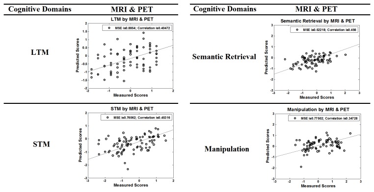
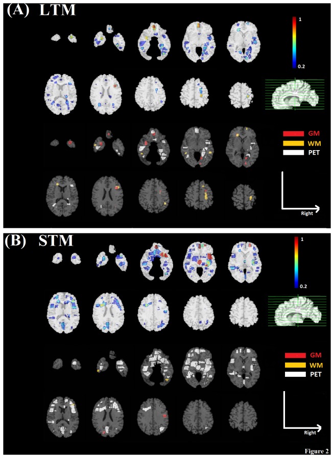
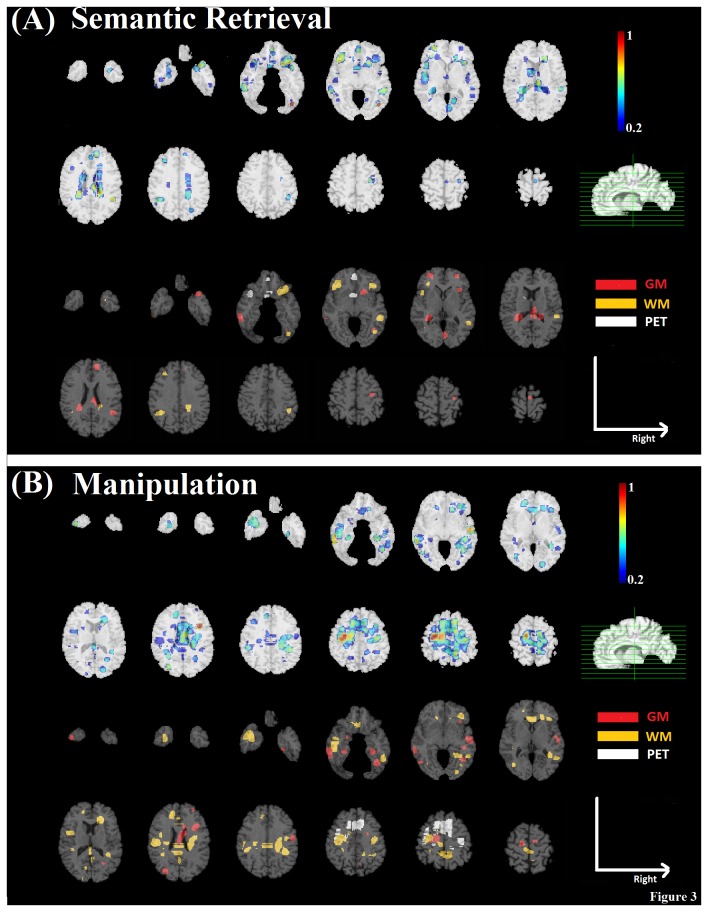
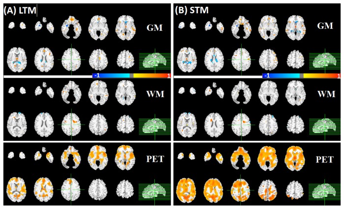
In this study, we used high-dimensional pattern regression methods based on structural (gray and white matter; GM and WM) and functional (positron emission tomography of regional cerebral blood flow; PET) brain data to identify cross-sectional imaging biomarkers of cognitive performance in cognitively normal older adults from the Baltimore Longitudinal Study of Aging (BLSA). We focused on specific components of executive and memory domains known to decline with aging, including manipulation, semantic retrieval, long-term memory (LTM), and short-term memory (STM). For each imaging modality, brain regions associated with each cognitive domain were generated by adaptive regional clustering. A relevance vector machine was adopted to model the nonlinear continuous relationship between brain regions and cognitive performance, with cross-validation to select the most informative brain regions (using recursive feature elimination) as imaging biomarkers and optimize model parameters. Predicted cognitive scores using our regression algorithm based on the resulting brain regions correlated well with actual performance. Also, regression models obtained using combined GM, WM, and PET imaging modalities outperformed models based on single modalities. Imaging biomarkers related to memory performance included the orbito-frontal and medial temporal cortical regions with LTM showing stronger correlation with the temporal lobe than STM. Brain regions predicting executive performance included orbito-frontal, and occipito-temporal areas. The PET modality had higher contribution to most cognitive domains except manipulation, which had higher WM contribution from the superior longitudinal fasciculus and the genu of the corpus callosum. These findings based on machine-learning methods demonstrate the importance of combining structural and functional imaging data in understanding complex cognitive mechanisms and also their potential usage as biomarkers that predict cognitive status.
在本研究中,我们使用基于大脑结构(灰质和白质;GM和WM)和功能(区域脑血流正电子发射断层扫描;PET)数据的高维模式回归方法,从巴尔的摩衰老纵向研究(BLSA)中识别认知正常的老年人认知表现的横断面成像生物标志物。我们聚焦于已知会随衰老而下降的执行和记忆领域的特定组成部分,包括操作、语义检索、长期记忆(LTM)和短期记忆(STM)。对于每种成像模态,通过自适应区域聚类生成与每个认知领域相关的脑区。采用相关向量机对脑区与认知表现之间的非线性连续关系进行建模,并通过交叉验证来选择信息量最大的脑区(使用递归特征消除)作为成像生物标志物,并优化模型参数。使用基于所得脑区的回归算法预测的认知分数与实际表现相关性良好。此外,使用GM、WM和PET成像模态组合获得的回归模型优于基于单一模态的模型。与记忆表现相关的成像生物标志物包括眶额和内侧颞叶皮质区域,LTM与颞叶的相关性比STM更强。预测执行表现的脑区包括眶额和枕颞区域。PET模态对大多数认知领域的贡献更高,但操作领域除外,操作领域中,上纵束和胼胝体膝部的WM贡献更高。这些基于机器学习方法的发现证明了在理解复杂认知机制中结合结构和功能成像数据的重要性,以及它们作为预测认知状态生物标志物的潜在用途。