Laboratory of Molecular Imaging and Nanomedicine, National Institute of Biomedical Imaging and Bioengineering , National Institutes of Health , Bethesda, Maryland 20892, United States.
J Am Chem Soc. 2014 Feb 5;136(5):1706-9. doi: 10.1021/ja410438n. Epub 2014 Jan 17.
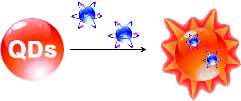
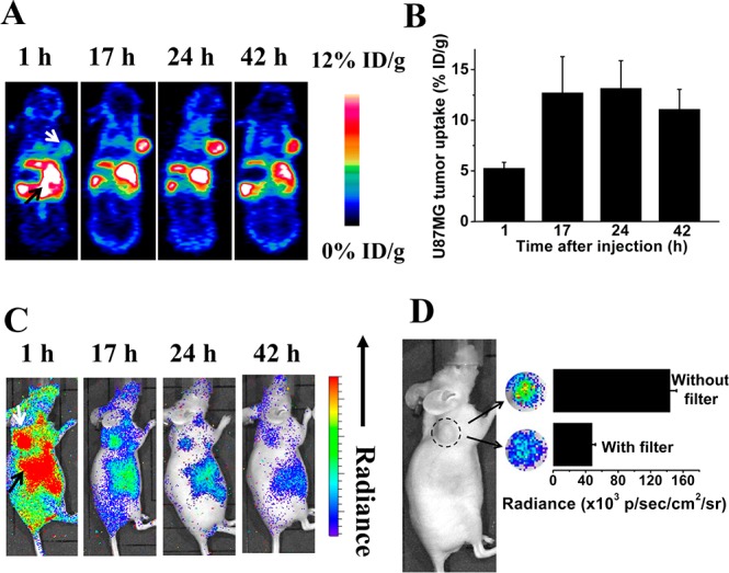
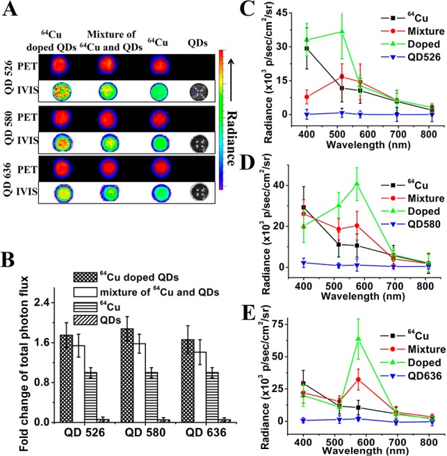
Construction of self-illuminating semiconducting nanocrystals, also called quantum dots (QDs), has attracted much attention recently due to their potential as highly sensitive optical probes for biological imaging applications. Here we prepared a self-illuminating QD system by doping positron-emitting radionuclide (64)Cu into CdSe/ZnS core/shell QDs via a cation-exchange reaction. The (64)Cu-doped CdSe/ZnS QDs exhibit efficient Cerenkov resonance energy transfer (CRET). The signal of (64)Cu can accurately reflect the biodistribution of the QDs during circulation with no dissociation of (64)Cu from the nanoparticles. We also explored this system for in vivo tumor imaging. This nanoprobe showed high tumor-targeting ability in a U87MG glioblastoma xenograft model (12.7% ID/g at 17 h time point) and feasibility for in vivo luminescence imaging of tumor in the absence of excitation light. The availability of these self-illuminating integrated QDs provides an accurate and convenient tool for in vivo tumor imaging and detection.
自发光半导体纳米晶体(也称为量子点,quantum dots,QDs)的构建由于其在生物成像应用中作为高灵敏度光学探针的潜力而受到广泛关注。在这里,我们通过阳离子交换反应将正电子发射放射性核素(64)Cu 掺杂到 CdSe/ZnS 核/壳 QD 中,制备了自发光 QD 系统。(64)Cu 掺杂的 CdSe/ZnS QD 表现出有效的契伦科夫共振能量转移(CRET)。信号(64)Cu 可以准确反映 QD 在循环过程中的生物分布,而不会从纳米颗粒中解离(64)Cu。我们还探索了该系统用于体内肿瘤成像。该纳米探针在 U87MG 胶质母细胞瘤异种移植模型中表现出高肿瘤靶向能力(17 h 时间点为 12.7% ID/g),并且在没有激发光的情况下可用于体内肿瘤的发光成像。这些自发光集成 QD 的可用性为体内肿瘤成像和检测提供了一种准确、方便的工具。