Van Houten Bennett, Kad Neil
Department of Pharmacology and Chemical Biology, University of Pittsburgh Cancer Institute, University of Pittsburgh, Pittsburgh, PA, USA.
School of Biological Sciences, University of Essex, Wivenhoe Park, Colchester CO4 3SQ, UK.
DNA Repair (Amst). 2014 Aug;20:41-48. doi: 10.1016/j.dnarep.2013.10.012. Epub 2014 Jan 25.
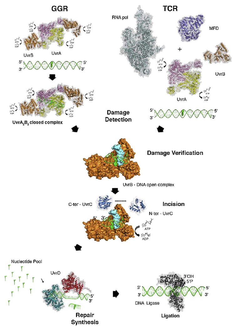
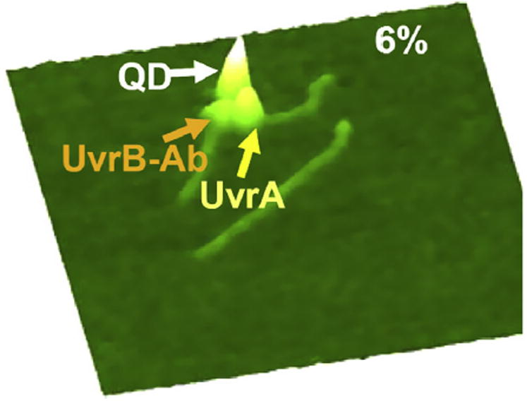
Despite three decades of biochemical and structural analysis of the prokaryotic nucleotide excision repair (NER) system, many intriguing questions remain with regard to how the UvrA, UvrB, and UvrC proteins detect, verify and remove a wide range of DNA lesions. Single-molecule techniques have begun to allow more detailed understanding of the kinetics and action mechanism of this complex process. This article reviews how atomic force microscopy and fluorescence microscopy have captured new glimpses of how these proteins work together to mediate NER.
尽管对原核生物核苷酸切除修复(NER)系统进行了三十年的生化和结构分析,但关于UvrA、UvrB和UvrC蛋白如何检测、验证和去除各种DNA损伤仍存在许多有趣的问题。单分子技术已开始使人们能够更详细地了解这一复杂过程的动力学和作用机制。本文综述了原子力显微镜和荧光显微镜如何为这些蛋白质协同介导NER的工作方式带来了新的认识。