• 文献检索
  • 文档翻译
  • 深度研究
  • 学术资讯
  • Suppr Zotero 插件Zotero 插件
  • 邀请有礼
  • 套餐&价格
  • 历史记录
应用&插件
Suppr Zotero 插件Zotero 插件浏览器插件Mac 客户端Windows 客户端微信小程序
定价
高级版会员购买积分包购买API积分包
服务
文献检索文档翻译深度研究API 文档MCP 服务
关于我们
关于 Suppr公司介绍联系我们用户协议隐私条款
关注我们

Suppr 超能文献

核心技术专利:CN118964589B侵权必究
粤ICP备2023148730 号-1Suppr @ 2026

文献检索

告别复杂PubMed语法,用中文像聊天一样搜索,搜遍4000万医学文献。AI智能推荐,让科研检索更轻松。

立即免费搜索

文件翻译

保留排版,准确专业,支持PDF/Word/PPT等文件格式,支持 12+语言互译。

免费翻译文档

深度研究

AI帮你快速写综述,25分钟生成高质量综述,智能提取关键信息,辅助科研写作。

立即免费体验

解析肾小球滤过屏障各组成部分的三维结构。

Resolution of the three dimensional structure of components of the glomerular filtration barrier.

机构信息

Nanoscale Physics Research Laboratories, School of Physics and Astronomy, University of Birmingham, Birmingham, UK.

出版信息

BMC Nephrol. 2014 Feb 1;15:24. doi: 10.1186/1471-2369-15-24.

DOI:10.1186/1471-2369-15-24
PMID:24484633
原文链接:https://pmc.ncbi.nlm.nih.gov/articles/PMC3922634/
Abstract

BACKGROUND

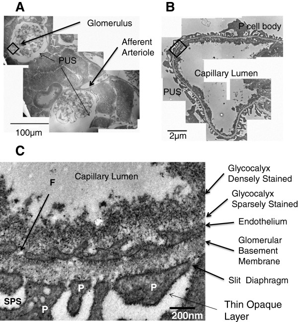
The human glomerulus is the primary filtration unit of the kidney, and contains the Glomerular Filtration Barrier (GFB). The GFB had been thought to comprise 3 layers - the endothelium, the basement membrane and the podocyte foot processes. However, recent studies have suggested that at least two additional layers contribute to the function of the GFB, the endothelial glycocalyx on the vascular side, and the sub-podocyte space on the urinary side. To investigate the structure of these additional layers is difficult as it requires three-dimensional reconstruction of delicate sub-microscopic (<1 μm) cellular and extracellular elements.

METHODS

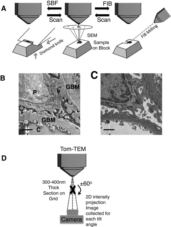
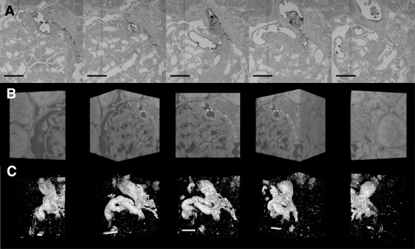
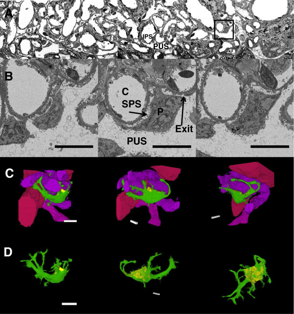
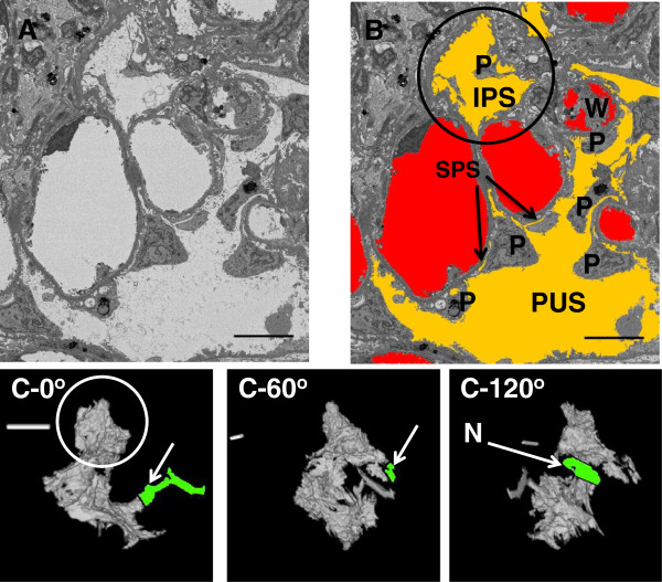
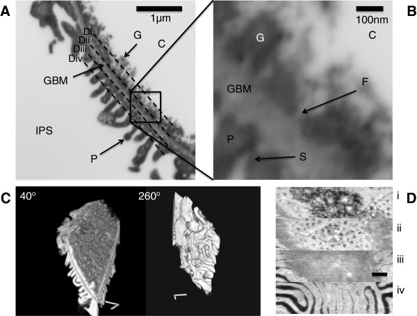
Here we have combined three different advanced electron microscopic techniques that cover multiple orders of magnitude of volume sampled, with a novel staining methodology (Lanthanum Dysprosium Glycosaminoglycan adhesion, or LaDy GAGa), to determine the structural basis of these two additional layers. Serial Block Face Scanning Electron Microscopy (SBF-SEM) was used to generate a 3-D image stack with a volume of a 5.3 x 105 μm3 volume of a whole kidney glomerulus (13% of glomerular volume). Secondly, Focused Ion Beam milling Scanning Electron Microscopy (FIB-SEM) was used to image a filtration region (48 μm3 volume). Lastly Transmission Electron Tomography (Tom-TEM) was performed on a 0.3 μm3 volume to identify the fine structure of the glycocalyx.

RESULTS

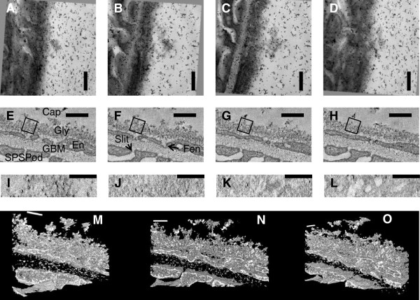
Tom-TEM clearly showed 20 nm fibre spacing in the glycocalyx, within a limited field of view. FIB-SEM demonstrated, in a far greater field of view, how the glycocalyx structure related to fenestrations and the filtration slits, though without the resolution of TomTEM. SBF-SEM was able to determine the extent of the sub-podocyte space and glycocalyx coverage, without additional heavy metal staining. Neither SBF- nor FIB-SEM suffered the anisotropic shrinkage under the electron beam that is seen with Tom-TEM.

CONCLUSIONS

These images demonstrate that the three dimensional structure of the GFB can be imaged, and investigated from the whole glomerulus to the fine structure of the glycocalyx using three dimensional electron microscopy techniques. This should allow the identification of structural features regulating physiology, and their disruption in pathological states, aiding the understanding of kidney disease.

摘要

背景

人类肾小球是肾脏的主要过滤单位,包含肾小球滤过屏障 (GFB)。GFB 曾被认为由 3 层组成 - 内皮细胞、基底膜和足细胞的足突。然而,最近的研究表明,至少还有另外两层有助于 GFB 的功能,即血管侧的内皮糖萼和尿侧的亚足突空间。研究这些额外层的结构非常困难,因为它需要对细微的亚微观 (<1μm) 细胞和细胞外元素进行三维重建。

方法

在这里,我们结合了三种不同的先进电子显微镜技术,这些技术涵盖了多个体积采样数量级,并采用了一种新的染色方法(镧钕糖胺聚糖粘连,或 LaDy GAGa),以确定这两个额外层的结构基础。连续块面扫描电子显微镜 (SBF-SEM) 用于生成一个 3-D 图像堆栈,其体积为整个肾脏肾小球的 5.3 x 105 μm3 体积(肾小球体积的 13%)。其次,聚焦离子束铣削扫描电子显微镜 (FIB-SEM) 用于对过滤区域进行成像(48 μm3 体积)。最后,在 0.3 μm3 体积上进行透射电子断层扫描 (Tom-TEM) 以识别糖萼的精细结构。

结果

Tom-TEM 清楚地显示了糖萼中的 20nm 纤维间距,在有限的视野内。FIB-SEM 在更大的视野中展示了糖萼结构如何与窗孔和过滤缝隙相关,尽管没有 TomTEM 的分辨率。SBF-SEM 能够确定亚足突空间和糖萼覆盖范围的程度,而无需额外的重金属染色。SBF- 和 FIB-SEM 都没有像 Tom-TEM 那样在电子束下出现各向异性收缩。

结论

这些图像表明,可以使用三维电子显微镜技术从整个肾小球到糖萼的精细结构来对 GFB 的三维结构进行成像和研究。这应该允许识别调节生理学的结构特征及其在病理状态下的破坏,从而有助于理解肾脏疾病。

https://cdn.ncbi.nlm.nih.gov/pmc/blobs/6f9a/3922634/0b148197e2d0/1471-2369-15-24-7.jpg
https://cdn.ncbi.nlm.nih.gov/pmc/blobs/6f9a/3922634/fb5d171e69e6/1471-2369-15-24-1.jpg
https://cdn.ncbi.nlm.nih.gov/pmc/blobs/6f9a/3922634/f4b5ba6e9570/1471-2369-15-24-2.jpg
https://cdn.ncbi.nlm.nih.gov/pmc/blobs/6f9a/3922634/e6780ac84345/1471-2369-15-24-3.jpg
https://cdn.ncbi.nlm.nih.gov/pmc/blobs/6f9a/3922634/1fb03c10190f/1471-2369-15-24-4.jpg
https://cdn.ncbi.nlm.nih.gov/pmc/blobs/6f9a/3922634/67db0159736a/1471-2369-15-24-5.jpg
https://cdn.ncbi.nlm.nih.gov/pmc/blobs/6f9a/3922634/8fda449af78f/1471-2369-15-24-6.jpg
https://cdn.ncbi.nlm.nih.gov/pmc/blobs/6f9a/3922634/0b148197e2d0/1471-2369-15-24-7.jpg

相似文献

1
Resolution of the three dimensional structure of components of the glomerular filtration barrier.解析肾小球滤过屏障各组成部分的三维结构。
BMC Nephrol. 2014 Feb 1;15:24. doi: 10.1186/1471-2369-15-24.
2
3D reconstruction of the glycocalyx structure in mammalian capillaries using electron tomography.使用电子断层扫描技术对哺乳动物毛细血管中糖萼结构进行三维重建。
Microcirculation. 2012 May;19(4):343-51. doi: 10.1111/j.1549-8719.2012.00168.x.
3
Sepsis induces albuminuria and alterations in the glomerular filtration barrier: a morphofunctional study in the rat.败血症导致蛋白尿和肾小球滤过屏障的改变:大鼠的形态功能研究。
Crit Care. 2011;15(6):R277. doi: 10.1186/cc10559. Epub 2011 Nov 22.
4
Combined Electron Microscopy Approaches for Arterial Glycocalyx Visualization.用于动脉糖萼可视化的联合电子显微镜方法
Front Cardiovasc Med. 2022 Mar 9;9:840689. doi: 10.3389/fcvm.2022.840689. eCollection 2022.
5
Loss of the Endothelial Glycocalyx Component EMCN Leads to Glomerular Impairment.内皮糖萼成分EMCN的缺失导致肾小球损伤。
Circ Res. 2025 Jan 3;136(1):59-74. doi: 10.1161/CIRCRESAHA.124.325218. Epub 2024 Nov 25.
6
Three-dimensional imaging of podocyte ultrastructure using FE-SEM and FIB-SEM tomography.使用 FE-SEM 和 FIB-SEM 断层扫描技术对足细胞超微结构进行三维成像。
Cell Tissue Res. 2020 Feb;379(2):245-254. doi: 10.1007/s00441-019-03118-3. Epub 2019 Nov 22.
7
Application of transmission electron tomography for modeling the renal corpuscle.应用透射电子断层成像术对肾小球进行建模。
Pathol Res Pract. 2013 Nov;209(11):731-4. doi: 10.1016/j.prp.2013.06.013. Epub 2013 Jul 8.
8
Combining serial block face and focused ion beam scanning electron microscopy for 3D studies of rare events.结合连续块面和聚焦离子束扫描电子显微镜用于罕见事件的三维研究。
Methods Cell Biol. 2019;152:87-101. doi: 10.1016/bs.mcb.2019.03.014. Epub 2019 May 7.
9
The role of the glomerular endothelium in albumin handling.肾小球内皮在白蛋白处理中的作用。
Nat Rev Nephrol. 2013 Dec;9(12):717-25. doi: 10.1038/nrneph.2013.197. Epub 2013 Oct 1.
10
3D imaging of the early embryonic chicken heart with focused ion beam scanning electron microscopy.利用聚焦离子束扫描电子显微镜对早期鸡胚心脏进行 3D 成像。
Microsc Microanal. 2014 Aug;20(4):1111-9. doi: 10.1017/S1431927614000828. Epub 2014 Apr 17.

引用本文的文献

1
Hemodynamic simulation and modeling of three-dimensional glomeruli at anatomical scale.解剖尺度下三维肾小球的血流动力学模拟与建模
Phys Fluids (1994). 2025 May;37(5):051907. doi: 10.1063/5.0264128. Epub 2025 May 16.
2
Factors influencing glycocalyx degradation: a narrative review.影响糖萼降解的因素:一项叙述性综述。
Front Immunol. 2025 Jan 16;15:1490395. doi: 10.3389/fimmu.2024.1490395. eCollection 2024.
3
An ultrathin membrane mediates tissue-specific morphogenesis and barrier function in a human kidney chip.超薄膜介导人类肾芯片中的组织特异性形态发生和屏障功能。

本文引用的文献

1
NIH Image to ImageJ: 25 years of image analysis.NIH 图像到 ImageJ:25 年的图像分析。
Nat Methods. 2012 Jul;9(7):671-5. doi: 10.1038/nmeth.2089.
2
Correlative light and volume electron microscopy: using focused ion beam scanning electron microscopy to image transient events in model organisms.相关光与体积电子显微镜技术:利用聚焦离子束扫描电子显微镜对模式生物中的瞬时事件进行成像。
Methods Cell Biol. 2012;111:357-82. doi: 10.1016/B978-0-12-416026-2.00018-2.
3
VEGF165b overexpression restores normal glomerular water permeability in VEGF164-overexpressing adult mice.
Sci Adv. 2024 Jun 7;10(23):eadn2689. doi: 10.1126/sciadv.adn2689. Epub 2024 Jun 5.
4
Destruction of vascular endothelial glycocalyx during formation of pre-metastatic niches.转移前生态位形成过程中血管内皮糖萼的破坏。
Heliyon. 2024 Apr 1;10(7):e29101. doi: 10.1016/j.heliyon.2024.e29101. eCollection 2024 Apr 15.
5
Modeling C3 glomerulopathies: C3 convertase regulation on an extracellular matrix surface.建模 C3 肾小球疾病:细胞外基质表面上的 C3 转化酶调节。
Front Immunol. 2023 Jan 18;13:1073802. doi: 10.3389/fimmu.2022.1073802. eCollection 2022.
6
Super-Resolution Microscopy: A Technique to Revolutionize Research and Diagnosis of Glomerulopathies.超分辨率显微镜:一种变革肾小球疾病研究与诊断的技术。
Glomerular Dis. 2022 Dec 18;3(1):19-28. doi: 10.1159/000528713. eCollection 2023 Jan-Dec.
7
Treatment of tubular damage in high-fat-diet-fed obese mice using sodium-glucose co-transporter inhibitors.使用钠-葡萄糖协同转运蛋白抑制剂治疗高脂肪饮食喂养肥胖小鼠的管状损伤。
PLoS One. 2023 Feb 13;18(2):e0281770. doi: 10.1371/journal.pone.0281770. eCollection 2023.
8
John squire and endothelial glycocalyx structure: an unfinished story.约翰·斯奎尔和内皮糖萼结构:一个未完成的故事。
J Muscle Res Cell Motil. 2023 Sep;44(3):217-223. doi: 10.1007/s10974-022-09629-x. Epub 2022 Oct 19.
9
Nondestructive cellular-level 3D observation of mouse kidney using laboratory-based X-ray microscopy with paraffin-mediated contrast enhancement.利用基于实验室的 X 射线显微镜和石蜡介导的对比增强,对小鼠肾脏进行无损细胞水平的 3D 观察。
Sci Rep. 2022 Jun 8;12(1):9436. doi: 10.1038/s41598-022-13394-9.
10
A Reinterpretation of Evidence for the Endothelial Glycocalyx Filtration Structure.对内皮糖萼过滤结构证据的重新诠释
Front Cell Dev Biol. 2021 Sep 1;9:734661. doi: 10.3389/fcell.2021.734661. eCollection 2021.
VEGF165b 的过表达可恢复过表达 VEGF164 的成年小鼠正常肾小球水通透性。
Am J Physiol Renal Physiol. 2012 Oct;303(7):F1026-36. doi: 10.1152/ajprenal.00410.2011. Epub 2012 Jul 18.
4
Loss of the endothelial glycocalyx links albuminuria and vascular dysfunction.内皮糖萼的丧失与蛋白尿和血管功能障碍有关。
J Am Soc Nephrol. 2012 Aug;23(8):1339-50. doi: 10.1681/ASN.2012010017. Epub 2012 Jul 12.
5
Fiji: an open-source platform for biological-image analysis.斐济:一个用于生物影像分析的开源平台。
Nat Methods. 2012 Jun 28;9(7):676-82. doi: 10.1038/nmeth.2019.
6
3D reconstruction of the glycocalyx structure in mammalian capillaries using electron tomography.使用电子断层扫描技术对哺乳动物毛细血管中糖萼结构进行三维重建。
Microcirculation. 2012 May;19(4):343-51. doi: 10.1111/j.1549-8719.2012.00168.x.
7
Similar endothelial glycocalyx structures in microvessels from a range of mammalian tissues: evidence for a common filtering mechanism?各种哺乳动物组织微血管中的类似内皮糖萼结构:是否存在共同的过滤机制?
Biophys J. 2011 Sep 7;101(5):1046-56. doi: 10.1016/j.bpj.2011.07.036.
8
Structure-function studies of blood and air capillaries in chicken lung using 3D electron microscopy.利用 3D 电子显微镜研究鸡肺中的血液和空气毛细血管的结构-功能。
Respir Physiol Neurobiol. 2010 Feb 28;170(2):202-9. doi: 10.1016/j.resp.2009.12.010. Epub 2009 Dec 28.
9
New aspects of glomerular filtration barrier structure and function: five layers (at least) not three.肾小球滤过屏障结构与功能的新观点:五层(至少)而非三层
Curr Opin Nephrol Hypertens. 2009 May;18(3):197-205. doi: 10.1097/MNH.0b013e328329f837.
10
Properties of the glomerular barrier and mechanisms of proteinuria.肾小球滤过屏障的特性及蛋白尿的机制。
Physiol Rev. 2008 Apr;88(2):451-87. doi: 10.1152/physrev.00055.2006.