Chevalier Laurence, Selim Jean, Castro Celia, Cuvilly Fabien, Baste Jean-Marc, Richard Vincent, Pareige Philippe, Bellien Jeremy
Université Rouen Normandie, CNRS, INSA Rouen Normandie- Normandie Université- GPM-UMR 6634, Rouen, France.
Université Rouen Normandie, INSERM, Normandie Université, ENVI- U1096, Rouen, France.
Front Cardiovasc Med. 2022 Mar 9;9:840689. doi: 10.3389/fcvm.2022.840689. eCollection 2022.
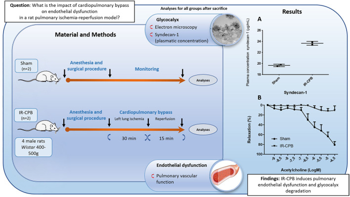
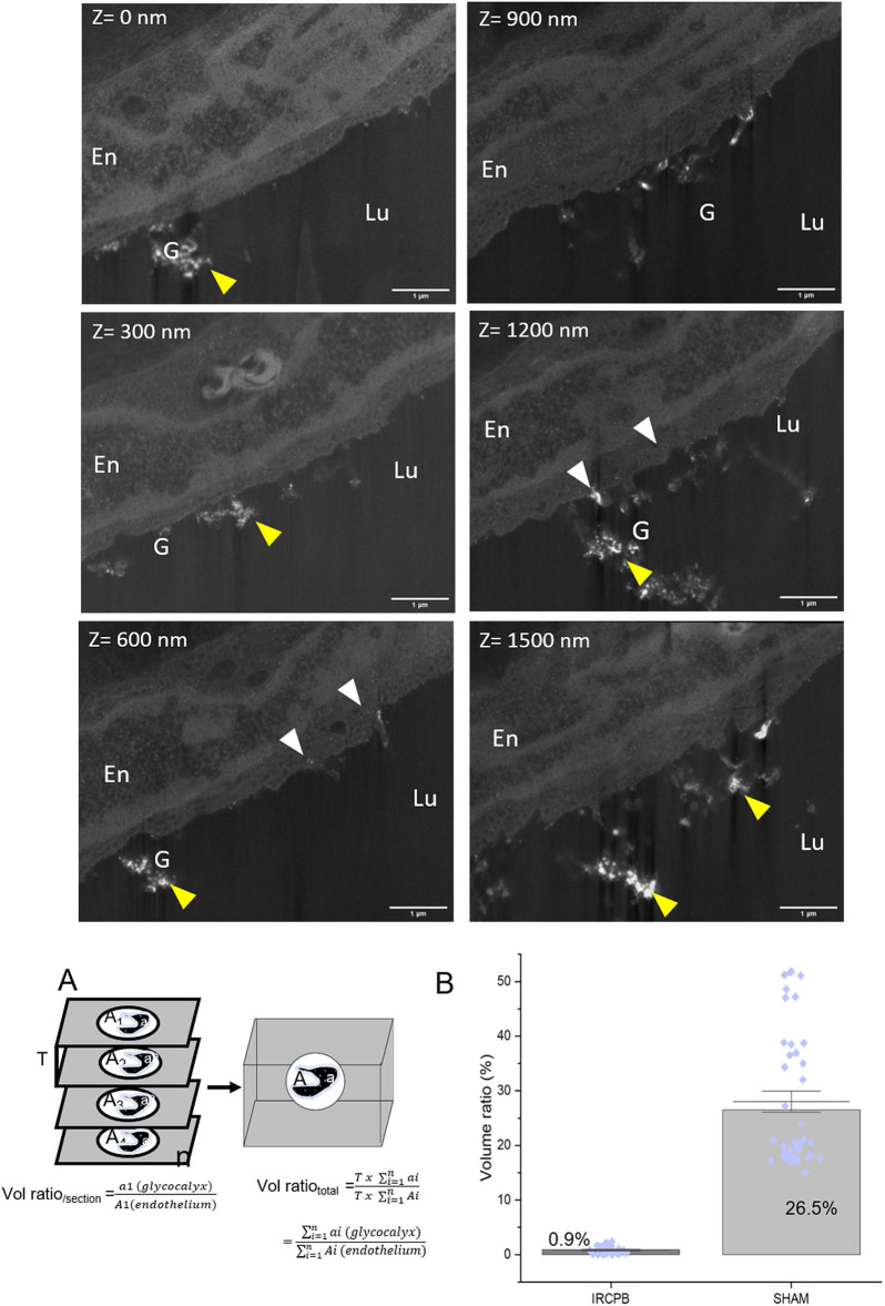
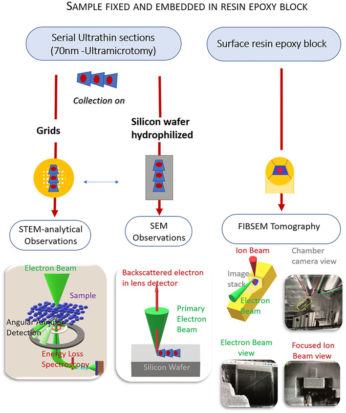
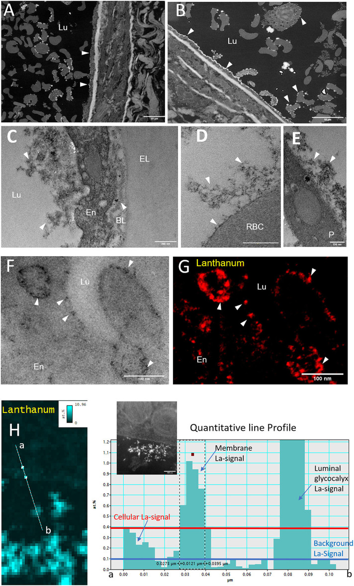
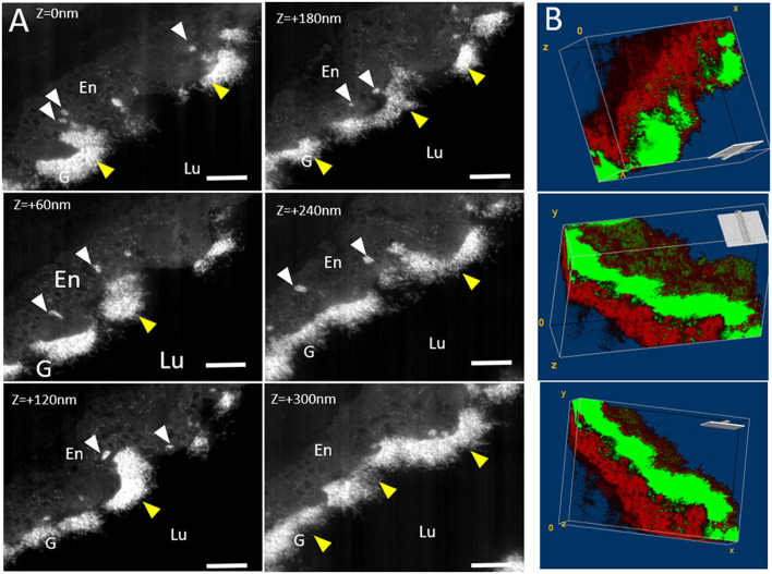
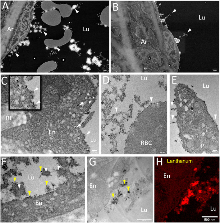
Mainly constituted of glycosaminoglycans and proteoglycans, the glycocalyx is anchored in the plasma membrane, covering, in particular, the extracellular face of the arterial endothelium. Due to its complex three-dimensional (3D) architecture, the glycocalyx interacts with a wide variety of proteins, contributing to vascular permeability, the flow of mechanotransduction, and the modulation of local inflammatory processes. Alterations of glycocalyx structure mediate the endothelial dysfunction and contribute to the aggravation of peripheral vascular diseases. Therefore, the exploration of its ultrastructure becomes a priority to evaluate the degree of injury under physiopathological conditions and to assess the impact of therapeutic approaches. The objective of this study was to develop innovative approaches in electron microscopy to visualize the glycocalyx at the subcellular scale. Intravenous perfusion on rats with a fixing solution containing aldehyde fixatives enriched with lanthanum ions was performed to prepare arterial samples. The addition of lanthanum nitrate in the fixing solution allowed the enhancement of the staining of the glycocalyx for transmission electron microscopy (TEM) and to detect elastic and inelastic scattered electrons, providing complementary qualitative information. The strength of scanning electron microscopy (SEM) was used on resin-embedded serial sections, allowing rapid and efficient large field imaging and previous correlative TEM observations for ultrastructural fine details. To demonstrate the dynamic feature of the glycocalyx, 3D tomography was provided by dual-beam focus-ion-beam-SEM (FIB-SEM). These approaches allowed us to visualize and characterize the ultrastructure of the pulmonary artery glycocalyx under physiological conditions and in a rat pulmonary ischemia-reperfusion model, known to induce endothelial dysfunction. This study demonstrates the feasibility of combined SEM, TEM, and FIB-SEM tomography approaches on the same sample as the multiscale visualization and the identification of structural indicators of arterial endothelial glycocalyx integrity.
糖萼主要由糖胺聚糖和蛋白聚糖组成,锚定在质膜中,尤其覆盖动脉内皮的细胞外表面。由于其复杂的三维(3D)结构,糖萼与多种蛋白质相互作用,有助于血管通透性、机械转导流以及局部炎症过程的调节。糖萼结构的改变介导内皮功能障碍,并导致外周血管疾病的加重。因此,探索其超微结构成为评估生理病理条件下损伤程度以及评估治疗方法影响的首要任务。本研究的目的是开发创新的电子显微镜方法,以在亚细胞尺度上可视化糖萼。对大鼠进行静脉灌注含富镧离子醛类固定剂的固定液,以制备动脉样本。在固定液中添加硝酸镧可增强用于透射电子显微镜(TEM)的糖萼染色,并检测弹性和非弹性散射电子,提供补充的定性信息。扫描电子显微镜(SEM)的优势用于树脂包埋的连续切片,可实现快速高效的大视野成像,并进行先前的相关TEM观察以获取超微结构细节。为了证明糖萼的动态特征,通过双束聚焦离子束扫描电子显微镜(FIB-SEM)提供三维断层扫描。这些方法使我们能够在生理条件下以及在已知会诱导内皮功能障碍的大鼠肺缺血再灌注模型中可视化和表征肺动脉糖萼的超微结构。本研究证明了在同一样本上结合SEM、TEM和FIB-SEM断层扫描方法进行多尺度可视化以及识别动脉内皮糖萼完整性结构指标的可行性。