Mehić Edin, Xu Julia M, Caler Connor J, Coulson Nathaniel K, Moritz Chet T, Mourad Pierre D
Department of Bioengineering, University of Washington, Seattle, Washington, United States of America.
Department of Materials Science and Engineering, University of Washington, Seattle, Washington, United States of America.
PLoS One. 2014 Feb 4;9(2):e86939. doi: 10.1371/journal.pone.0086939. eCollection 2014.
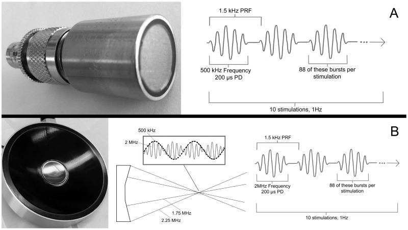
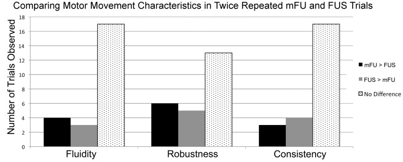
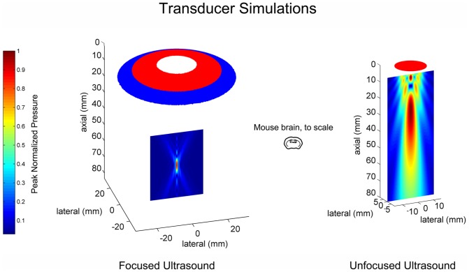
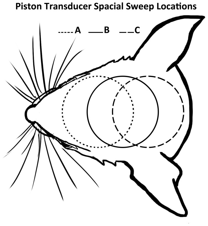
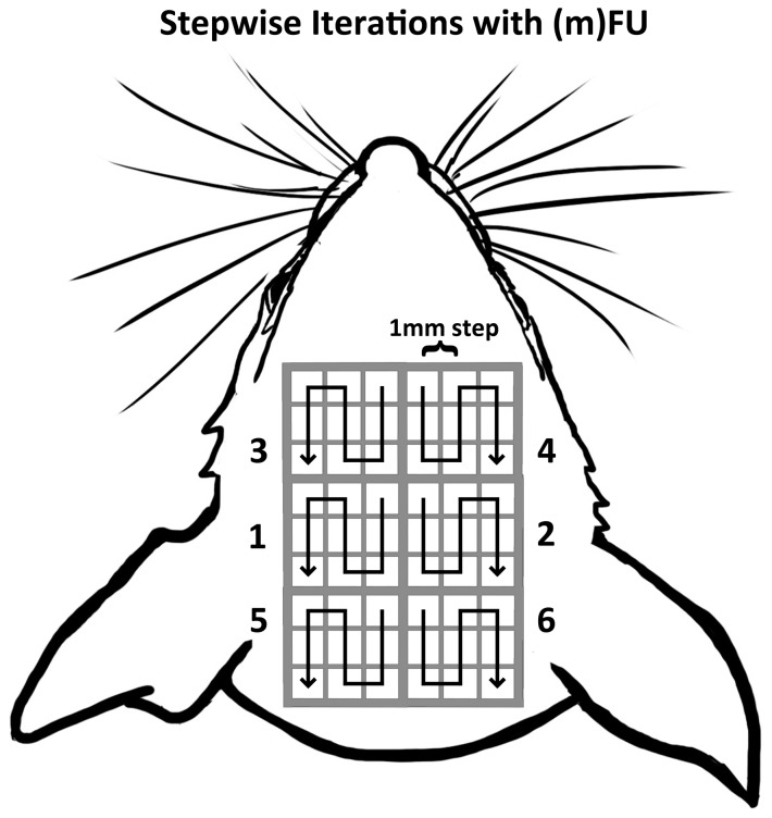
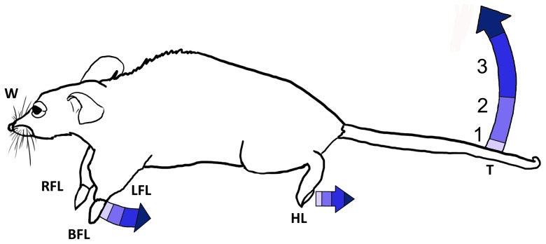
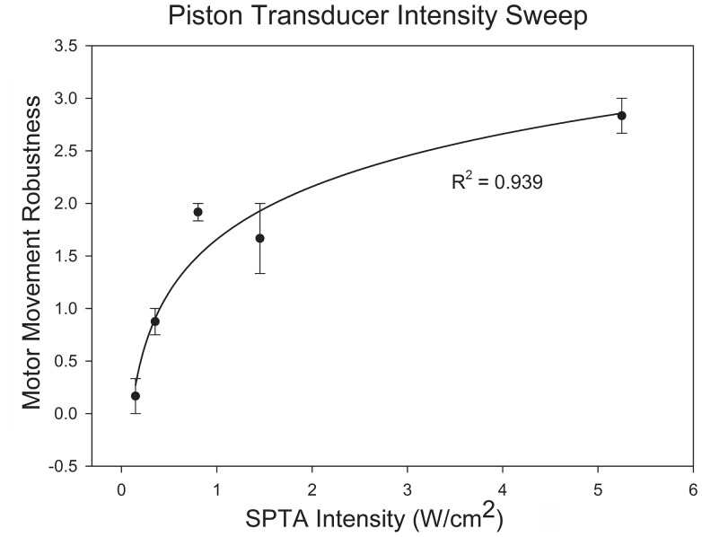
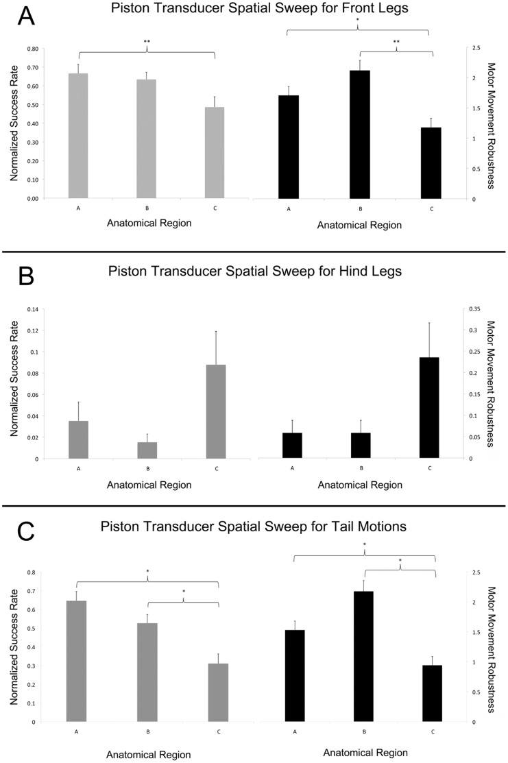
Transcranial ultrasound can alter brain function transiently and nondestructively, offering a new tool to study brain function now and inform future therapies. Previous research on neuromodulation implemented pulsed low-frequency (250-700 kHz) ultrasound with spatial peak temporal average intensities (ISPTA) of 0.1-10 W/cm(2). That work used transducers that either insonified relatively large volumes of mouse brain (several mL) with relatively low-frequency ultrasound and produced bilateral motor responses, or relatively small volumes of brain (on the order of 0.06 mL) with relatively high-frequency ultrasound that produced unilateral motor responses. This study seeks to increase anatomical specificity to neuromodulation with modulated focused ultrasound (mFU). Here, 'modulated' means modifying a focused 2-MHz carrier signal dynamically with a 500-kHz signal as in vibro-acoustography, thereby creating a low-frequency but small volume (approximately 0.015 mL) source of neuromodulation. Application of transcranial mFU to lightly anesthetized mice produced various motor movements with high spatial selectivity (on the order of 1 mm) that scaled with the temporal average ultrasound intensity. Alone, mFU and focused ultrasound (FUS) each induced motor activity, including unilateral motions, though anatomical location and type of motion varied. Future work should include larger animal models to determine the relative efficacy of mFU versus FUS. Other studies should determine the biophysical processes through which they act. Also of interest is exploration of the potential research and clinical applications for targeted, transcranial neuromodulation created by modulated focused ultrasound, especially mFU's ability to produce compact sources of ultrasound at the very low frequencies (10-100s of Hertz) that are commensurate with the natural frequencies of the brain.
经颅超声可以暂时且无损地改变脑功能,为当下研究脑功能及指导未来治疗提供了一种新工具。此前关于神经调节的研究采用了脉冲低频(250 - 700千赫)超声,空间峰值时间平均强度(ISPTA)为0.1 - 10瓦/平方厘米。该研究使用的换能器,要么以相对低频的超声照射相对大体积的小鼠脑(几毫升)并产生双侧运动反应,要么以相对高频的超声照射相对小体积的脑(约0.06毫升)并产生单侧运动反应。本研究旨在通过调制聚焦超声(mFU)提高神经调节的解剖学特异性。在此,“调制”是指像在振动声成像中那样,用500千赫的信号动态调制2兆赫的聚焦载波信号,从而产生一个低频但小体积(约0.015毫升)的神经调节源。将经颅mFU应用于轻度麻醉的小鼠,产生了具有高空间选择性(约1毫米量级)的各种运动,这些运动与时间平均超声强度成比例。单独使用时,mFU和聚焦超声(FUS)各自都能诱发运动活动,包括单侧运动,不过运动的解剖位置和类型有所不同。未来的工作应包括使用更大的动物模型来确定mFU与FUS的相对疗效。其他研究应确定它们起作用的生物物理过程。同样值得关注的是探索由调制聚焦超声产生的靶向经颅神经调节的潜在研究和临床应用,特别是mFU在与脑自然频率相当的极低频率(10 - 100赫兹)下产生紧凑超声源的能力。