Haritonova Alyona, Liu Dalong, Ebbini Emad S
IEEE Trans Ultrason Ferroelectr Freq Control. 2015 Dec;62(12):2031-42. doi: 10.1109/TUFFC.2014.006882.
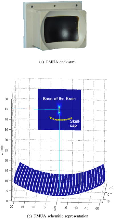
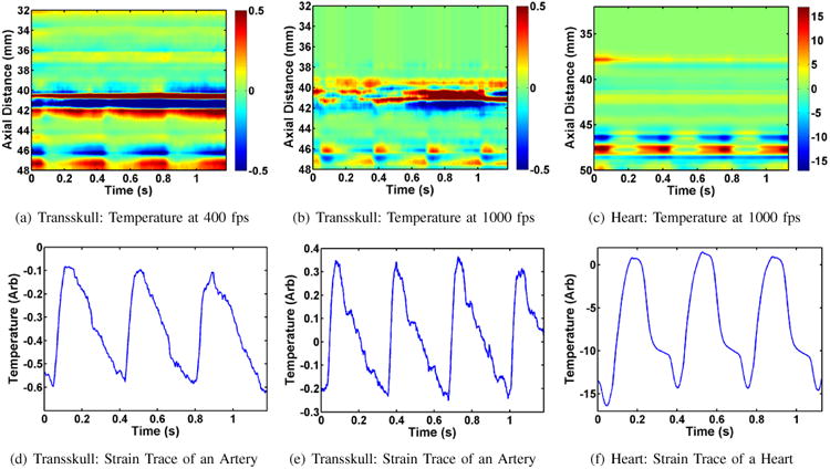
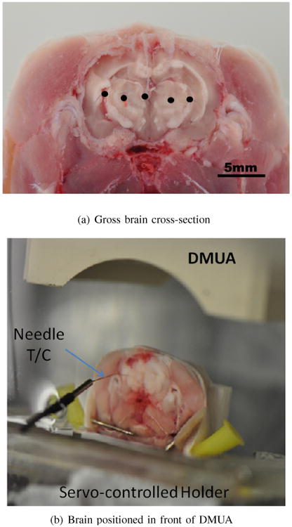
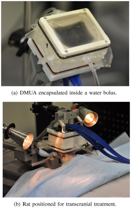
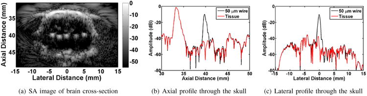
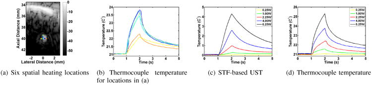
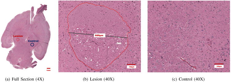
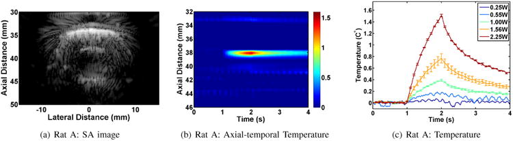
Focused ultrasound (FUS) has been proposed for a variety of transcranial applications, including neuromodulation, tumor ablation, and blood-brain barrier opening. A flurry of activity in recent years has generated encouraging results demonstrating its feasibility in these and other applications. To date, monitoring of FUS beams has been primarily accomplished using MR guidance, where both MR thermography and elastography have been used. The recent introduction of real-time dual-mode ultrasound array (DMUA) systems offers a new paradigm in transcranial focusing. In this paper, we present first experimental results of ultrasound-guided transcranial FUS (tFUS) application in a rodent brain, both ex vivo and in vivo. DMUA imaging is used for visualization of the treatment region for placement of the focal spot within the brain. This includes the detection and localization of pulsating blood vessels at or near the target point(s). In addition, DMUA imaging is used to monitor and localize the FUS-tissue interactions in real time. In particular, a concave (40 mm radius of curvature), 32-element, 3.5-MHz DMUA prototype was used for imaging and tFUS application in ex vivo and in vivo rat models. The ex vivo experiments were used to evaluate the point spread function of the transcranial DMUA imaging at various points within the brain. In addition, DMUA-based transcranial ultrasound thermography measurements were compared with thermocouple measurements of subtherapeutic tFUS heating in rat brain ex vivo. The ex vivo setting was also used to demonstrate the capability of DMUA to produce localized thermal lesions. The in vivo experiments were designed to demonstrate the ability of the DMUA to apply, monitor, and localize subtherapeutic tFUS patterns that could be beneficial in transient blood-brain barrier opening. The results show that although the DMUA focus is degraded due to the propagation through the skull, it still produces localized heating effects within a sub-millimeter volume. In addition, DMUA transcranial echo data from brain tissue allow for reliable estimation of temperature change.
聚焦超声(FUS)已被应用于多种经颅治疗,包括神经调节、肿瘤消融和血脑屏障开放。近年来的一系列研究取得了令人鼓舞的成果,证明了其在这些及其他应用中的可行性。迄今为止,FUS束的监测主要通过磁共振(MR)引导完成,其中MR热成像和弹性成像均有应用。实时双模超声阵列(DMUA)系统的最新引入为经颅聚焦提供了一种新的模式。在本文中,我们展示了超声引导下经颅FUS(tFUS)在啮齿动物脑内离体和活体实验中的初步结果。DMUA成像用于可视化治疗区域,以便将焦点放置在脑内。这包括检测和定位靶点处或其附近的搏动血管。此外,DMUA成像用于实时监测和定位FUS与组织的相互作用。具体而言,一个曲率半径为40mm的凹面、32阵元、3.5MHz的DMUA原型被用于离体和活体大鼠模型的成像及tFUS应用。离体实验用于评估经颅DMUA成像在脑内不同点的点扩散函数。此外,还将基于DMUA的经颅超声热成像测量结果与大鼠脑内离体亚治疗性tFUS加热的热电偶测量结果进行了比较。离体实验还用于证明DMUA产生局部热损伤的能力。活体实验旨在证明DMUA应用、监测和定位亚治疗性tFUS模式的能力,这些模式可能有助于短暂性血脑屏障开放。结果表明,尽管DMUA焦点在穿过颅骨传播时会退化,但它仍能在亚毫米体积内产生局部加热效应。此外,来自脑组织的DMUA经颅回波数据能够可靠地估计温度变化。