Institute of Physical Chemistry "Rocasolano". CSIC. Madrid, Spain.
Carlos III Hospital, Immunology Department, Madrid, Spain.
PLoS Negl Trop Dis. 2014 Mar 6;8(3):e2735. doi: 10.1371/journal.pntd.0002735. eCollection 2014 Mar.
Anisakiasis is a re-emerging global disease caused by consumption of raw or lightly cooked fish contaminated with L3 Anisakis larvae. This zoonotic disease is characterized by severe gastrointestinal and/or allergic symptoms which may misdiagnosed as appendicitis, gastric ulcer or other food allergies. The Anisakis allergen Ani s 5 is a protein belonging to the SXP/RAL-2 family; it is detected exclusively in nematodes. Previous studies showed that SXP/RAL-2 proteins are active antigens; however, their structure and function remain unknown. The aim of this study was to elucidate the three-dimensional structure of Ani s 5 and its main IgE and IgG4 binding regions.
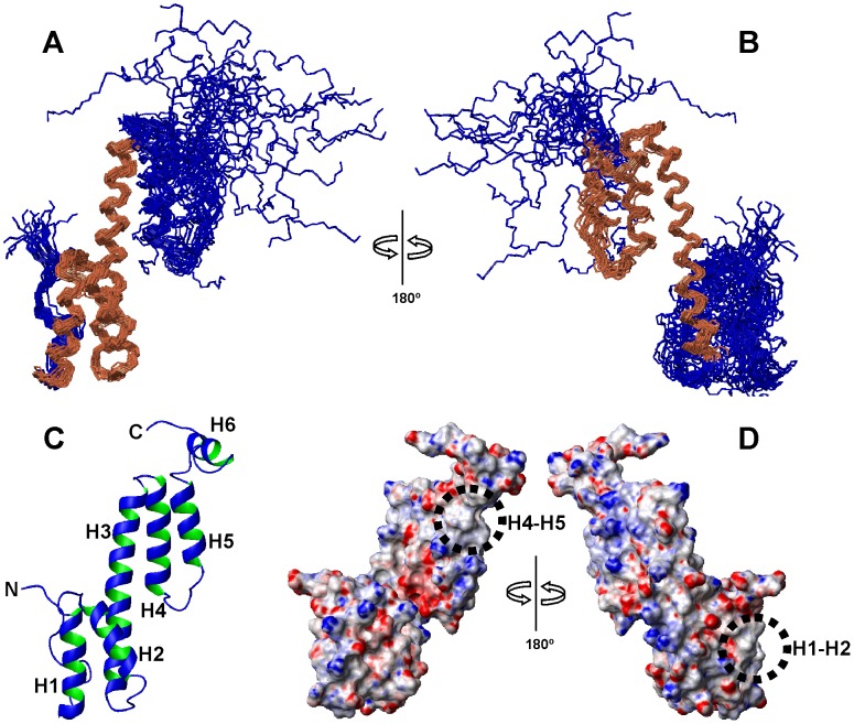
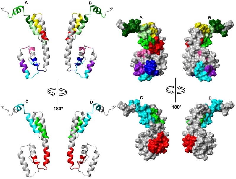
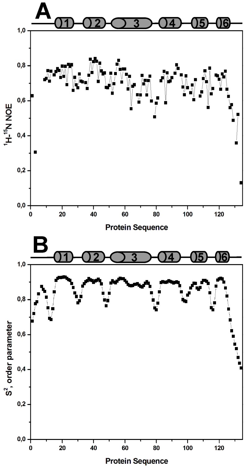
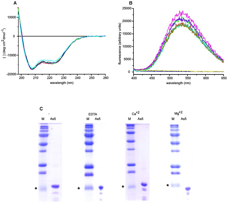
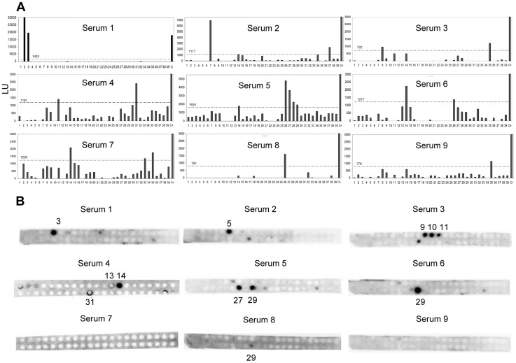
METHODOLOGY/PRINCIPAL FINDINGS: The tertiary structure of recombinant Ani s 5 in solution was solved by nuclear magnetic resonance. Mg2+, but not Ca2+, binding was determined by band shift using SDS-PAGE. IgE and IgG4 epitopes were elucidated by microarray immunoassay and SPOTs membranes using sera from nine Anisakis allergic patients. The tertiary structure of Ani s 5 is composed of six alpha helices (H), with a Calmodulin like fold. H3 is a long, central helix that organizes the structure, with H1 and H2 packing at its N-terminus and H4 and H5 packing at its C-terminus. The orientation of H6 is undefined. Regarding epitopes recognized by IgE and IgG4 immunoglobulins, the same eleven peptides derived from Ani s 5 were bound by both IgE and IgG4. Peptides 14 (L40-K59), 26 (A76-A95) and 35 (I103-D122) were recognized by three out of nine sera.
CONCLUSIONS/SIGNIFICANCE: This is the first reported 3D structure of an Anisakis allergen. Magnesium ion binding and structural resemblance to Calmodulin, suggest some putative functions for SXP/RAL-2 proteins. Furthermore, the IgE/IgG4 binding regions of Ani s 5 were identified as segments localized on its surface. These data will contribute towards a better understanding of the interactions that occur between immunoglobulins and allergens and, in turn, facilitate the design of novel diagnostic tests and immunotherapeutic strategies.
异尖线虫病是一种由食用受 L3 异尖线虫幼虫污染的生的或轻度熟的鱼引起的新兴全球性疾病。这种人畜共患疾病的特征是严重的胃肠道和/或过敏症状,可能误诊为阑尾炎、胃溃疡或其他食物过敏。异尖线虫过敏原 Ani s 5 是一种属于 SXP/RAL-2 家族的蛋白质;它仅在线虫中被检测到。先前的研究表明,SXP/RAL-2 蛋白是活性抗原;然而,它们的结构和功能仍然未知。本研究旨在阐明 Ani s 5 的三维结构及其主要 IgE 和 IgG4 结合区域。
方法/主要发现:通过核磁共振解决了重组 Ani s 5 在溶液中的三级结构。通过 SDS-PAGE 中的带位移确定 Mg2+,而不是 Ca2+的结合。使用来自 9 名异尖线虫过敏患者的血清通过微阵列免疫测定和 SPOTs 膜阐明 IgE 和 IgG4 表位。Ani s 5 的三级结构由六个α螺旋(H)组成,具有钙调蛋白样折叠。H3 是一个长的中央螺旋,组织结构,H1 和 H2 在其 N 末端包装,H4 和 H5 在其 C 末端包装。H6 的方向未定义。关于 IgE 和 IgG4 免疫球蛋白识别的表位,来自 Ani s 5 的相同的 11 个肽都被 IgE 和 IgG4 结合。肽 14(L40-K59)、26(A76-A95)和 35(I103-D122)被 9 份血清中的 3 份识别。
结论/意义:这是首例报道的异尖线虫过敏原的 3D 结构。镁离子结合和与钙调蛋白的结构相似性表明 SXP/RAL-2 蛋白具有某些推测的功能。此外,Ani s 5 的 IgE/IgG4 结合区域被鉴定为位于其表面的片段。这些数据将有助于更好地理解免疫球蛋白和过敏原之间发生的相互作用,并进而促进新型诊断测试和免疫治疗策略的设计。