• 文献检索
  • 文档翻译
  • 深度研究
  • 学术资讯
  • Suppr Zotero 插件Zotero 插件
  • 邀请有礼
  • 套餐&价格
  • 历史记录
应用&插件
Suppr Zotero 插件Zotero 插件浏览器插件Mac 客户端Windows 客户端微信小程序
定价
高级版会员购买积分包购买API积分包
服务
文献检索文档翻译深度研究API 文档MCP 服务
关于我们
关于 Suppr公司介绍联系我们用户协议隐私条款
关注我们

Suppr 超能文献

核心技术专利:CN118964589B侵权必究
粤ICP备2023148730 号-1Suppr @ 2026

文献检索

告别复杂PubMed语法,用中文像聊天一样搜索,搜遍4000万医学文献。AI智能推荐,让科研检索更轻松。

立即免费搜索

文件翻译

保留排版,准确专业,支持PDF/Word/PPT等文件格式,支持 12+语言互译。

免费翻译文档

深度研究

AI帮你快速写综述,25分钟生成高质量综述,智能提取关键信息,辅助科研写作。

立即免费体验

相似文献

1
In vitro evaluation of carbon-nanotube-reinforced bioprintable vascular conduits.碳纳米管增强型可生物打印血管导管的体外评估
Nanotechnology. 2014 Apr 11;25(14):145101. doi: 10.1088/0957-4484/25/14/145101. Epub 2014 Mar 14.
2
Effect of multiwall carbon nanotube reinforcement on coaxially extruded cellular vascular conduits.多壁碳纳米管增强对同轴挤压多孔血管导管的影响。
Mater Sci Eng C Mater Biol Appl. 2014 Jun 1;39:126-33. doi: 10.1016/j.msec.2014.02.036. Epub 2014 Feb 24.
3
UV-Assisted 3D Bioprinting of Nanoreinforced Hybrid Cardiac Patch for Myocardial Tissue Engineering.基于紫外光辅助 3D 生物打印的纳米增强型心脏组织工程复合补片
Tissue Eng Part C Methods. 2018 Feb;24(2):74-88. doi: 10.1089/ten.TEC.2017.0346. Epub 2017 Nov 30.
4
Moldable elastomeric polyester-carbon nanotube scaffolds for cardiac tissue engineering.用于心脏组织工程的可模塑弹性体聚酯-碳纳米管支架
Acta Biomater. 2017 Apr 1;52:81-91. doi: 10.1016/j.actbio.2016.12.009. Epub 2016 Dec 8.
5
In Vitro Study of Directly Bioprinted Perfusable Vasculature Conduits.直接生物打印可灌注血管导管的体外研究
Biomater Sci. 2015 Jan;3(1):134-43. doi: 10.1039/C4BM00234B.
6
Carbon nanotube reinforced hybrid microgels as scaffold materials for cell encapsulation.碳纳米管增强杂化微凝胶作为细胞包封的支架材料。
ACS Nano. 2012 Jan 24;6(1):362-72. doi: 10.1021/nn203711s. Epub 2011 Dec 20.
7
Carbon nanotubes in scaffolds for tissue engineering.用于组织工程的支架中的碳纳米管。
Expert Rev Med Devices. 2009 Sep;6(5):499-505. doi: 10.1586/erd.09.29.
8
Smooth muscle alpha-actin and calponin expression and extracellular matrix production of human coronary artery smooth muscle cells in 3D scaffolds.人冠状动脉平滑肌细胞在三维支架中的平滑肌α-肌动蛋白和钙调蛋白表达及细胞外基质生成
Tissue Eng Part A. 2009 Oct;15(10):3001-11. doi: 10.1089/ten.TEA.2009.0057.
9
Functionalized carbon nanotube reinforced scaffolds for bone regenerative engineering: fabrication, in vitro and in vivo evaluation.用于骨再生工程的功能化碳纳米管增强支架:制备、体外和体内评价
Biomed Mater. 2014 Jun;9(3):035001. doi: 10.1088/1748-6041/9/3/035001. Epub 2014 Mar 31.
10
Dielectrophoretically aligned carbon nanotubes to control electrical and mechanical properties of hydrogels to fabricate contractile muscle myofibers.电泳排列的碳纳米管控制水凝胶的电学和力学性能,以制造可收缩的肌肉肌纤维。
Adv Mater. 2013 Aug 7;25(29):4028-34. doi: 10.1002/adma.201301300. Epub 2013 Jun 25.

引用本文的文献

1
Fibroblast proximity to a tumor impacts fibroblast extracellular vesicles produced by 3D bioprinted stromal models.成纤维细胞与肿瘤的接近程度会影响由3D生物打印基质模型产生的成纤维细胞细胞外囊泡。
Biomater Sci. 2025 Jun 10. doi: 10.1039/d4bm01569j.
2
State of the Art of Bioengineering Approaches in Beta-Cell Replacement.β细胞替代中生物工程方法的现状
Curr Transplant Rep. 2025;12(1):17. doi: 10.1007/s40472-025-00470-y. Epub 2025 May 6.
3
Recent Advances in Hydrogel-Based 3D Bioprinting and Its Potential Application in the Treatment of Congenital Heart Disease.水凝胶基 3D 生物打印的最新进展及其在先天性心脏病治疗中的潜在应用。
Biomolecules. 2024 Jul 18;14(7):861. doi: 10.3390/biom14070861.
4
Gelation of Uniform Interfacial Diffusant in Embedded 3D Printing.嵌入式3D打印中均匀界面扩散剂的凝胶化
Adv Funct Mater. 2023 Dec 8;33(50). doi: 10.1002/adfm.202307435. Epub 2023 Aug 1.
5
Highly oriented hydrogels for tissue regeneration: design strategies, cellular mechanisms, and biomedical applications.用于组织再生的高度取向水凝胶:设计策略、细胞机制和生物医学应用。
Theranostics. 2024 Feb 24;14(5):1982-2035. doi: 10.7150/thno.89493. eCollection 2024.
6
Diffusion-Based 3D Bioprinting Strategies.基于扩散的 3D 生物打印策略。
Adv Sci (Weinh). 2024 Feb;11(8):e2306470. doi: 10.1002/advs.202306470. Epub 2023 Dec 25.
7
Nanomaterials-incorporated hydrogels for 3D bioprinting technology.用于3D生物打印技术的纳米材料复合水凝胶
Nano Converg. 2023 Nov 15;10(1):52. doi: 10.1186/s40580-023-00402-5.
8
Additive manufacturing of sustainable biomaterials for biomedical applications.用于生物医学应用的可持续生物材料的增材制造。
Asian J Pharm Sci. 2023 May;18(3):100812. doi: 10.1016/j.ajps.2023.100812. Epub 2023 Apr 27.
9
Manufacturing the multiscale vascular hierarchy: progress toward solving the grand challenge of tissue engineering.制造多尺度血管层次结构:解决组织工程重大挑战的进展。
Trends Biotechnol. 2023 Nov;41(11):1400-1416. doi: 10.1016/j.tibtech.2023.04.003. Epub 2023 May 9.
10
Block Copolymers in 3D/4D Printing: Advances and Applications as Biomaterials.3D/4D打印中的嵌段共聚物:作为生物材料的进展与应用
Polymers (Basel). 2023 Jan 8;15(2):322. doi: 10.3390/polym15020322.

本文引用的文献

1
Current approaches to electrospun nanofibers for tissue engineering.用于组织工程的静电纺丝纳米纤维的当前方法。
Biomed Mater. 2013 Feb;8(1):014102. doi: 10.1088/1748-6041/8/1/014102.
2
Characterization of printable cellular micro-fluidic channels for tissue engineering.用于组织工程的可打印细胞微流控通道的特性描述。
Biofabrication. 2013 Jun;5(2):025004. doi: 10.1088/1758-5082/5/2/025004. Epub 2013 Mar 5.
3
3D hybrid wound devices for spatiotemporally controlled release kinetics.用于时空控制释放动力学的 3D 混合伤口装置。
Comput Methods Programs Biomed. 2012 Dec;108(3):922-31. doi: 10.1016/j.cmpb.2012.05.004. Epub 2012 Jun 4.
4
Mechanical properties and in vitro behavior of nanofiber-hydrogel composites for tissue engineering applications.用于组织工程应用的纳米纤维-水凝胶复合材料的力学性能及体外行为。
Nanotechnology. 2012 Mar 9;23(9):095705. doi: 10.1088/0957-4484/23/9/095705. Epub 2012 Feb 10.
5
Chemically Functionalized Carbon Nanotubes as Substrates for Neuronal Growth.化学功能化碳纳米管作为神经元生长的基质
Nano Lett. 2004 Mar;4(3):507-511. doi: 10.1021/nl035193d.
6
Modeling the controllable pH-responsive swelling and pore size of networked alginate based biomaterials.模拟基于海藻酸盐的网络化生物材料的可控pH响应性溶胀和孔径。
Biomaterials. 2009 Oct;30(30):6119-29. doi: 10.1016/j.biomaterials.2009.07.034. Epub 2009 Aug 5.
7
Influence of surface oxides on the colloidal stability of multi-walled carbon nanotubes: a structure-property relationship.表面氧化物对多壁碳纳米管胶体稳定性的影响:结构-性能关系
Langmuir. 2009 Sep 1;25(17):9767-76. doi: 10.1021/la901128k.
8
Fabrication, characterization, and biocompatibility of single-walled carbon nanotube-reinforced alginate composite scaffolds manufactured using freeform fabrication technique.使用自由成型制造技术制备的单壁碳纳米管增强藻酸盐复合支架的制造、表征及生物相容性
J Biomed Mater Res B Appl Biomater. 2008 Nov;87(2):406-14. doi: 10.1002/jbm.b.31118.
9
Alginate hydrogels as biomaterials.藻酸盐水凝胶作为生物材料。
Macromol Biosci. 2006 Aug 7;6(8):623-33. doi: 10.1002/mabi.200600069.
10
Cellular toxicity of carbon-based nanomaterials.碳基纳米材料的细胞毒性
Nano Lett. 2006 Jun;6(6):1121-5. doi: 10.1021/nl060162e.

碳纳米管增强型可生物打印血管导管的体外评估

In vitro evaluation of carbon-nanotube-reinforced bioprintable vascular conduits.

作者信息

Dolati Farzaneh, Yu Yin, Zhang Yahui, De Jesus Aribet M, Sander Edward A, Ozbolat Ibrahim T

机构信息

Biomanufacturing Laboratory, Center for Computer-Aided Design, The University of Iowa, Iowa City, IA, USA. Mechanical and Industrial Engineering Department, The University of Iowa, Iowa City, IA, USA.

出版信息

Nanotechnology. 2014 Apr 11;25(14):145101. doi: 10.1088/0957-4484/25/14/145101. Epub 2014 Mar 14.

DOI:10.1088/0957-4484/25/14/145101
PMID:24632802
原文链接:https://pmc.ncbi.nlm.nih.gov/articles/PMC4281171/
Abstract

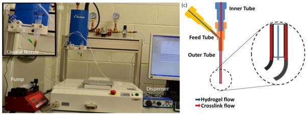
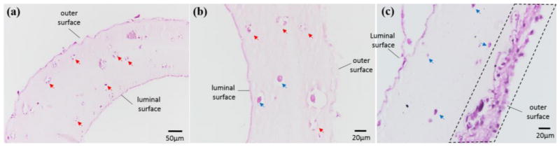
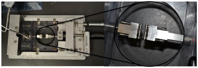
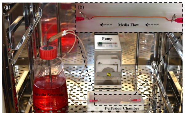
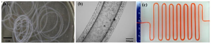
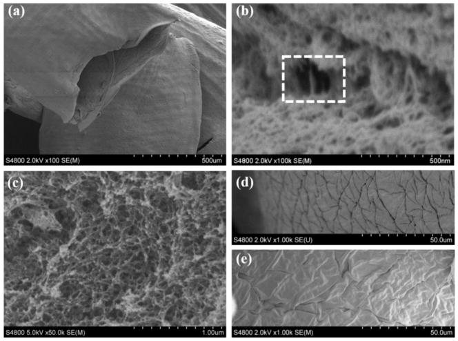
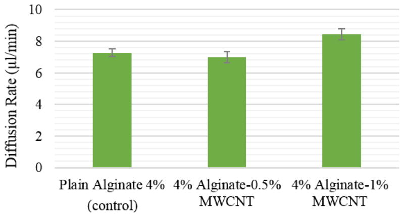
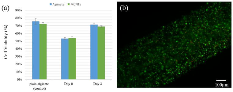
Vascularization of thick engineered tissue and organ constructs like the heart, liver, pancreas or kidney remains a major challenge in tissue engineering. Vascularization is needed to supply oxygen and nutrients and remove waste in living tissues and organs through a network that should possess high perfusion ability and significant mechanical strength and elasticity. In this paper, we introduce a fabrication process to print vascular conduits directly, where conduits were reinforced with carbon nanotubes (CNTs) to enhance their mechanical properties and bioprintability. In vitro evaluation of printed conduits encapsulated in human coronary artery smooth muscle cells was performed to characterize the effects of CNT reinforcement on the mechanical, perfusion and biological performance of the conduits. Perfusion and permeability, cell viability, extracellular matrix formation and tissue histology were assessed and discussed, and it was concluded that CNT-reinforced vascular conduits provided a foundation for mechanically appealing constructs where CNTs could be replaced with natural protein nanofibers for further integration of these conduits in large-scale tissue fabrication.

摘要

对于心脏、肝脏、胰腺或肾脏等厚实的工程组织和器官构建体而言,血管化仍然是组织工程中的一项重大挑战。血管化是通过一个应具备高灌注能力、显著机械强度和弹性的网络,来为活组织和器官提供氧气和营养物质,并清除废物。在本文中,我们介绍了一种直接打印血管导管的制造工艺,其中导管用碳纳米管(CNT)进行增强,以提高其机械性能和生物打印性。对包裹在人冠状动脉平滑肌细胞中的打印导管进行了体外评估,以表征碳纳米管增强对导管机械、灌注和生物学性能的影响。评估并讨论了灌注与渗透性、细胞活力、细胞外基质形成和组织病理学,得出的结论是,碳纳米管增强的血管导管为具有机械吸引力的构建体奠定了基础,在大规模组织制造中,可以用天然蛋白质纳米纤维替代碳纳米管,以进一步整合这些导管。

https://cdn.ncbi.nlm.nih.gov/pmc/blobs/7c9b/4281171/edb481c00717/nihms576705f9.jpg
https://cdn.ncbi.nlm.nih.gov/pmc/blobs/7c9b/4281171/c6d8fa07e1a0/nihms576705f1.jpg
https://cdn.ncbi.nlm.nih.gov/pmc/blobs/7c9b/4281171/6955f5b0e23a/nihms576705f2.jpg
https://cdn.ncbi.nlm.nih.gov/pmc/blobs/7c9b/4281171/2531a7d2bb39/nihms576705f3.jpg
https://cdn.ncbi.nlm.nih.gov/pmc/blobs/7c9b/4281171/654c7c609c79/nihms576705f4.jpg
https://cdn.ncbi.nlm.nih.gov/pmc/blobs/7c9b/4281171/946fc1b91d31/nihms576705f5.jpg
https://cdn.ncbi.nlm.nih.gov/pmc/blobs/7c9b/4281171/86ec8de852c9/nihms576705f6.jpg
https://cdn.ncbi.nlm.nih.gov/pmc/blobs/7c9b/4281171/0f594888a3a2/nihms576705f7.jpg
https://cdn.ncbi.nlm.nih.gov/pmc/blobs/7c9b/4281171/68f00523babb/nihms576705f8.jpg
https://cdn.ncbi.nlm.nih.gov/pmc/blobs/7c9b/4281171/edb481c00717/nihms576705f9.jpg