Zhou Jianhong, Han Yong, Lu Shemin
State Key Laboratory for Mechanical Behavior of Materials, Xi'an Jiaotong University, Xi'an, People's Republic of China.
Department of Genetics and Molecular Biology, College of Medicine, Xi'an Jiaotong University, Xi'an, People's Republic of China.
Int J Nanomedicine. 2014 Mar 8;9:1243-60. doi: 10.2147/IJN.S58236. eCollection 2014.
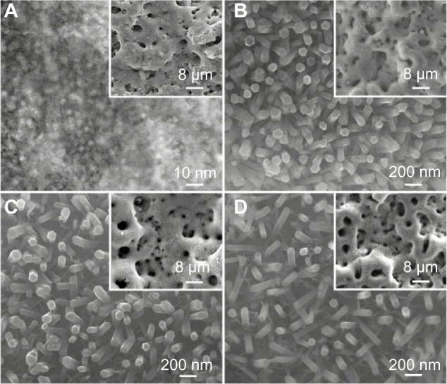
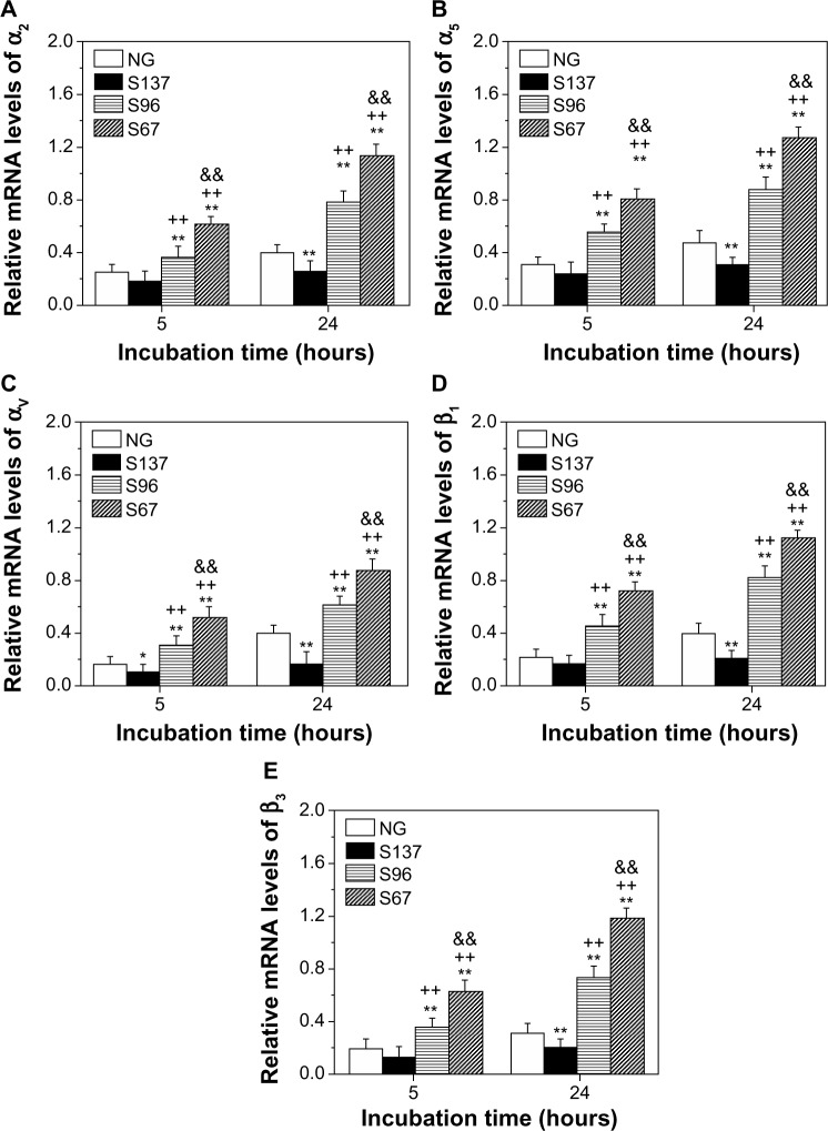
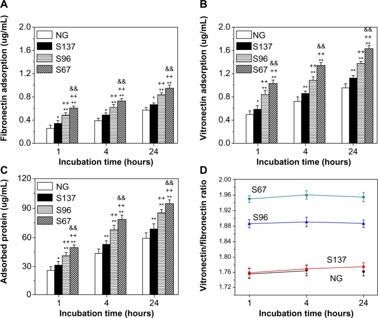
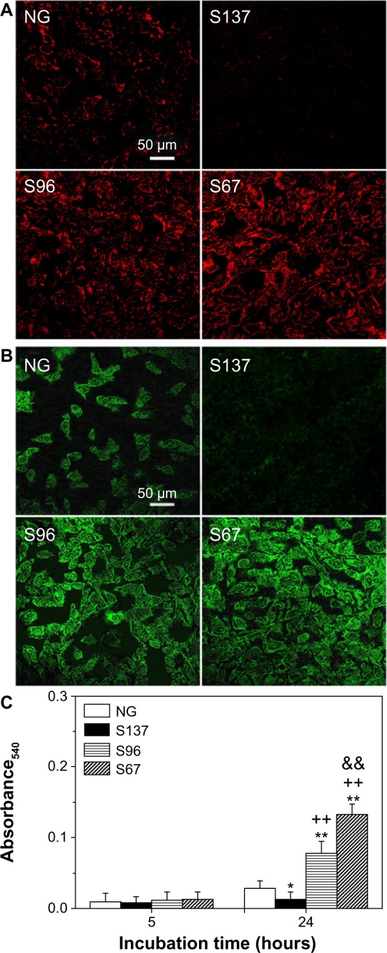
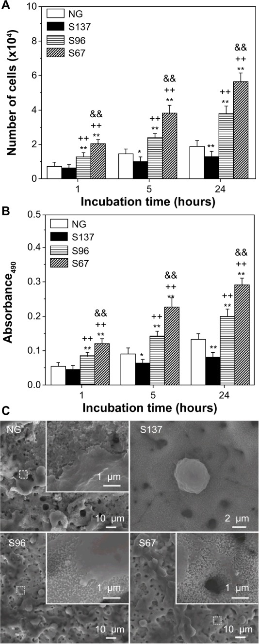
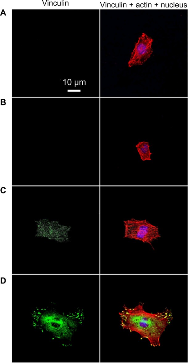
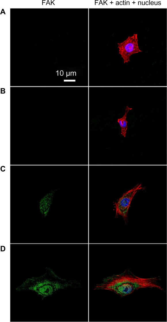
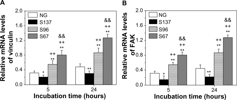
The process in which nanostructured surfaces mediate cell adhesion is not well understood, and may be indirect (via protein adsorption) or direct. We prepared Sr-doped hydroxyapatite (Sr1-HA) 3D nanorods (with interrod spacing of 67.3 ± 3.8, 95.7 ± 4.2, and 136.8 ± 8.7 nm) and 2D nanogranulate patterned coatings on titanium. Employing the coatings under the same surface chemistry and roughness, we investigated the indirect/direct role of Sr1-HA nanotopographies in the regulation of osteoblast adhesion in both serum-free and serum-containing Dulbecco's Modified Eagle/Ham's Medium. The results reveal that the number of adherent cells, cell-secreted anchoring proteins (fibronectin, vitronectin, and collagen), vinculin and focal adhesion kinase (FAK) denoted focal adhesion (FA) contact, and gene expression of vinculin, FAK, and integrin subunits (α2, α5, αv, β1, and β3), undergo significant changes in the inter-nanorod spacing and dimensionality of Sr1-HA nanotopographies in the absence of serum; they are significantly enhanced on the <96 nm spaced nanorods and more pronounced with decreasing interrod spacing. However, they are inhibited on the >96 nm spaced nanorods compared to nanogranulated 2D topography. Although the adsorption of fibronectin and vitronectin from serum are higher on 136.8 ± 8.7 nm spaced nanorod patterned topography than nanogranulated topography, cell adhesion is inhibited on the former compared to the latter in the presence of serum, further suggesting that reduced cell adhesion is independent of protein adsorption. It is clearly indicated that 3D nanotopography can directly modulate cell adhesion by regulating integrins, which subsequently mediate anchoring proteins' secretion and FA formation rather than via protein adsorption.
纳米结构表面介导细胞黏附的过程尚未完全明确,可能是间接的(通过蛋白质吸附)或直接的。我们制备了掺锶羟基磷灰石(Sr1-HA)三维纳米棒(棒间间距分别为67.3±3.8、95.7±4.2和136.8±8.7纳米)以及钛表面的二维纳米颗粒图案涂层。在相同的表面化学性质和粗糙度条件下使用这些涂层,我们研究了Sr1-HA纳米拓扑结构在无血清和含血清的杜氏改良 Eagle/Ham's培养基中对成骨细胞黏附调节的间接/直接作用。结果表明,在无血清条件下,黏附细胞数量、细胞分泌的锚定蛋白(纤连蛋白、玻连蛋白和胶原蛋白)、纽蛋白和粘着斑激酶(FAK)所代表的粘着斑(FA)接触,以及纽蛋白、FAK和整合素亚基(α2、α5、αv、β1和β3)的基因表达,在Sr1-HA纳米拓扑结构的纳米棒间距和维度上发生了显著变化;在间距<96纳米的纳米棒上显著增强,且随着棒间间距减小更为明显。然而,与纳米颗粒二维拓扑结构相比,在间距>96纳米的纳米棒上这些指标受到抑制。尽管血清中的纤连蛋白和玻连蛋白在间距为136.8±8.7纳米的纳米棒图案拓扑结构上的吸附高于纳米颗粒拓扑结构,但在有血清存在时,前者的细胞黏附却比后者受到抑制,这进一步表明细胞黏附减少与蛋白质吸附无关。显然,三维纳米拓扑结构可通过调节整合素来直接调节细胞黏附,随后介导锚定蛋白的分泌和粘着斑形成,而非通过蛋白质吸附。