Wang Junhua, Lin Renyong, Zhang Wenbao, Li Liang, Gottstein Bruno, Blagosklonov Oleg, Lü Guodong, Zhang Chuangshan, Lu Xiaomei, Vuitton Dominique A, Wen Hao
State Key Lab Incubation Base for Xinjiang Major Diseases Research and Xinjiang Key Laboratory of Echinococcosis, First Affiliated Hospital of Xinjiang Medical University, Urumqi, Xinjiang, China; Department of Nuclear Medicine, University of Franche-Comté and Jean Minjoz University Hospital, Besançon, Franche-Comté, France; Institute of Parasitology, University of Bern, Bern, Switzerland.
State Key Lab Incubation Base for Xinjiang Major Diseases Research and Xinjiang Key Laboratory of Echinococcosis, First Affiliated Hospital of Xinjiang Medical University, Urumqi, Xinjiang, China.
PLoS One. 2014 Mar 17;9(3):e91638. doi: 10.1371/journal.pone.0091638. eCollection 2014.
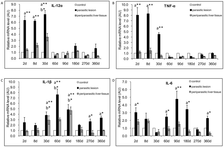
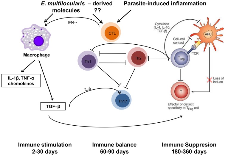
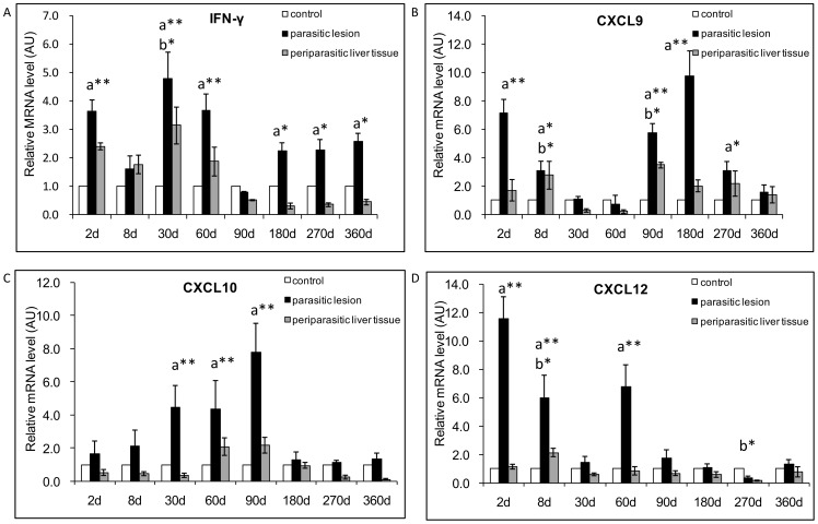
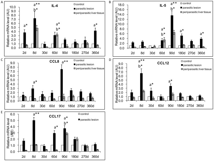
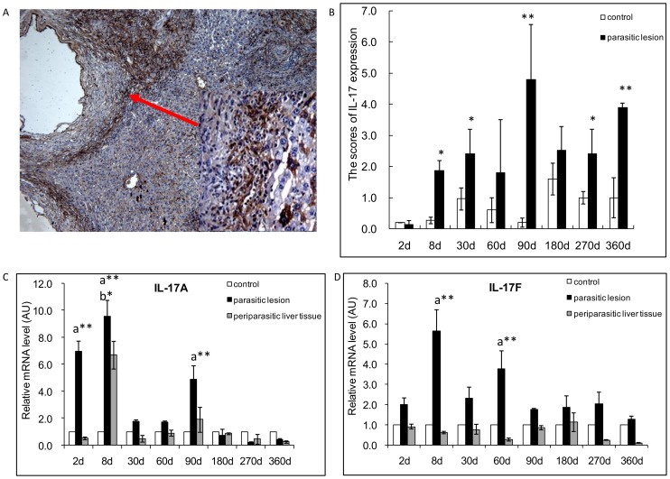
Pathogenesis of chronically developing alveolar echinococcosis (AE) is characterized by a continuous, granulomatous, periparasitic infiltration of immune cells surrounding the metacestode of Echinococcus multilocularis (E.multilocularis) in the affected liver. A detailed cytokine and chemokine profile analysis of the periparasitic infiltrate in the liver has, however, not yet been carried out in a comprehensive way all along the whole course of infection in E. multilocularis intermediate hosts. We thus assessed the hepatic gene expression profiles of 18 selected cytokine and chemokine genes using qRT-PCR in the periparasitic immune reaction and the subsequent adjacent, not directly affected, liver tissue of mice from day 2 to day 360 post intra-hepatic injection of metacestode. DNA microarray analysis was also used to get a more complete picture of the transcriptional changes occurring in the liver surrounding the parasitic lesions. Profiles of mRNA expression levels in the hepatic parasitic lesions showed that a mixed Th1/Th2 immune response, characterized by the concomitant presence of IL-12α, IFN-γ and IL-4, was established very early in the development of E. multilocularis. Subsequently, the profile extended to a combined tolerogenic profile associating IL-5, IL-10 and TGF-β. IL-17 was permanently expressed in the liver, mostly in the periparasitic infiltrate; this was confirmed by the increased mRNA expression of both IL-17A and IL-17F from a very early stage, with a subsequent decrease of IL-17A after this first initial rise. All measured chemokines were significantly expressed at a given stage of infection; their expression paralleled that of the corresponding Th1, Th2 or Th17 cytokines. In addition to giving a comprehensive insight in the time course of cytokines and chemokines in E. multilocularis lesion, this study contributes to identify new targets for possible immune therapy to minimize E. multilocularis-related pathology and to complement the only parasitostatic effect of benzimidazoles in AE.
慢性发展的肺泡型棘球蚴病(AE)的发病机制的特点是,在受影响的肝脏中,免疫细胞围绕多房棘球绦虫(E.multilocularis)的原头蚴持续进行肉芽肿性的、寄生虫周围浸润。然而,在多房棘球绦虫中间宿主感染的整个过程中,尚未全面开展对肝脏中寄生虫周围浸润物的细胞因子和趋化因子谱的详细分析。因此,我们通过qRT-PCR评估了肝内注射原头蚴后第2天至第360天小鼠的寄生虫周围免疫反应以及随后相邻的、未直接受影响的肝脏组织中18种选定的细胞因子和趋化因子基因的肝脏基因表达谱。还使用DNA微阵列分析以更全面地了解寄生虫病变周围肝脏中发生的转录变化。肝脏寄生虫病变中的mRNA表达水平谱显示,在多房棘球绦虫发育的早期就建立了以IL-12α、IFN-γ和IL-4同时存在为特征的混合Th1/Th2免疫反应。随后,该谱扩展为与IL-5、IL-10和TGF-β相关的联合耐受性谱。IL-17在肝脏中持续表达,主要在寄生虫周围浸润物中;这从很早阶段就通过IL-17A和IL-17F的mRNA表达增加得到证实,在首次初始升高后IL-17A随后下降。所有测定的趋化因子在感染的特定阶段均有显著表达;它们的表达与相应的Th1、Th2或Th17细胞因子的表达平行。除了全面深入了解多房棘球绦虫病变中细胞因子和趋化因子的时间进程外,本研究有助于确定可能的免疫治疗的新靶点,以尽量减少多房棘球绦虫相关的病理变化,并补充苯并咪唑在AE中唯一的抗寄生虫作用。