1] Department of Neurobiology, Stanford University School of Medicine, Stanford, California 94305, USA [2] Howard Hughes Medical Institute, Stanford University School of Medicine, Stanford, California 94305, USA.
Department of Neurobiology, Stanford University School of Medicine, Stanford, California 94305, USA.
Nature. 2014 Mar 27;507(7493):504-7. doi: 10.1038/nature13149.
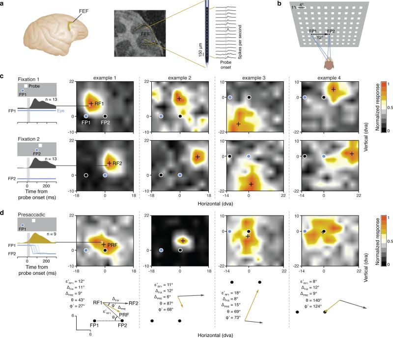
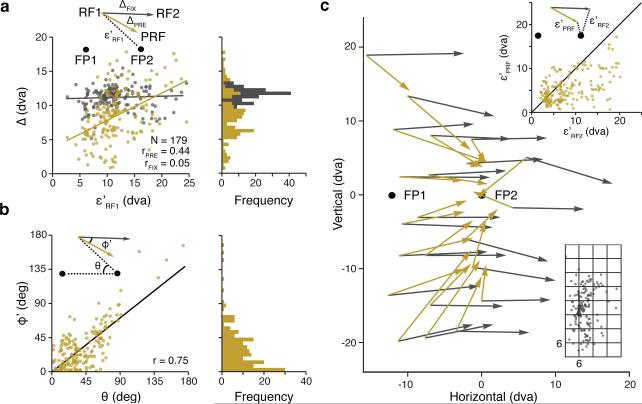
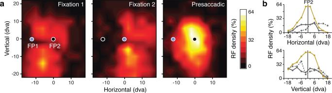
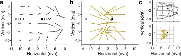
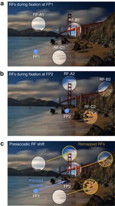
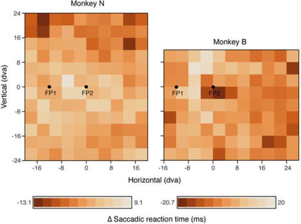
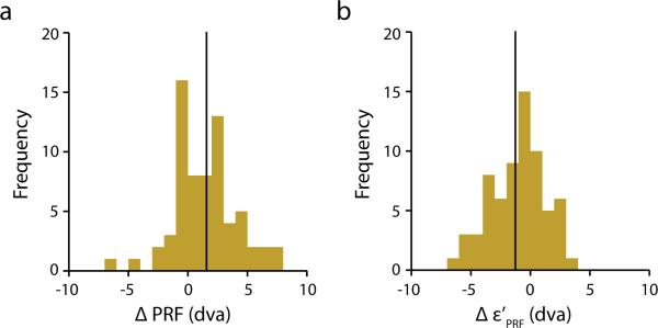
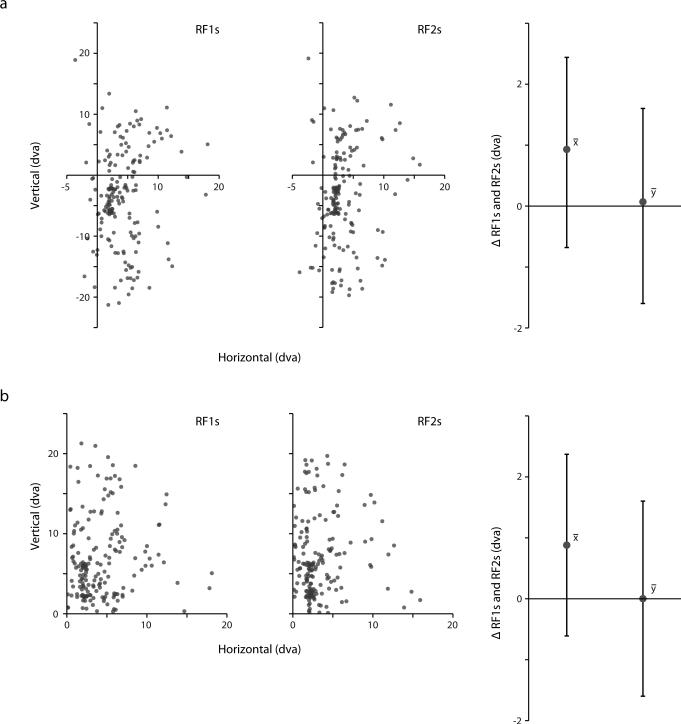
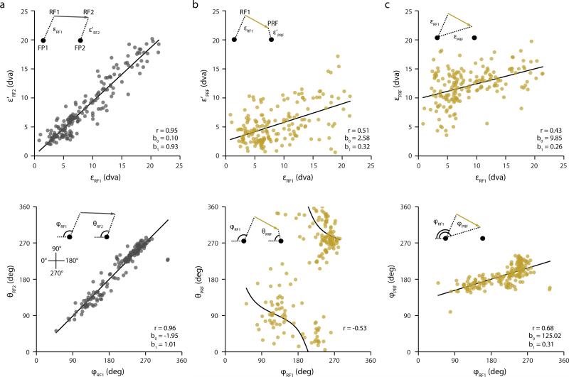
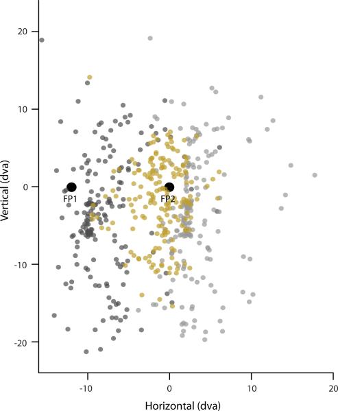
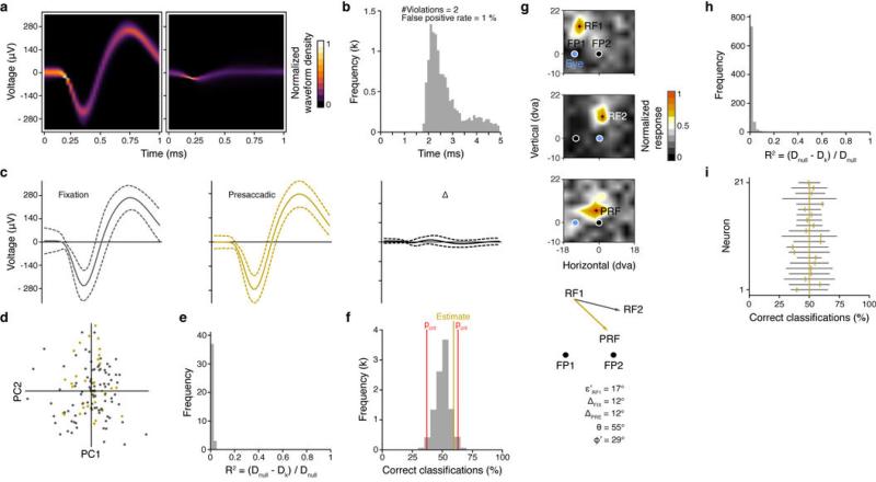
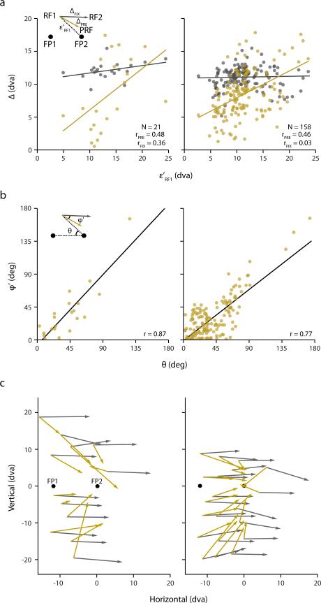
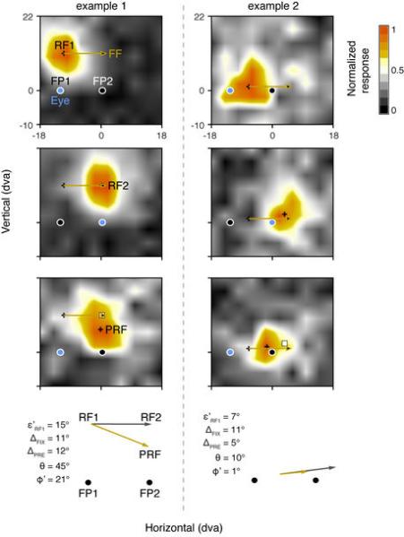
We experience the visual world through a series of saccadic eye movements, each one shifting our gaze to bring objects of interest to the fovea for further processing. Although such movements lead to frequent and substantial displacements of the retinal image, these displacements go unnoticed. It is widely assumed that a primary mechanism underlying this apparent stability is an anticipatory shifting of visual receptive fields (RFs) from their presaccadic to their postsaccadic locations before movement onset. Evidence of this predictive 'remapping' of RFs has been particularly apparent within brain structures involved in gaze control. However, critically absent among that evidence are detailed measurements of visual RFs before movement onset. Here we show that during saccade preparation, rather than remap, RFs of neurons in a prefrontal gaze control area massively converge towards the saccadic target. We mapped the visual RFs of prefrontal neurons during stable fixation and immediately before the onset of eye movements, using multi-electrode recordings in monkeys. Following movements from an initial fixation point to a target, RFs remained stationary in retinocentric space. However, in the period immediately before movement onset, RFs shifted by as much as 18 degrees of visual angle, and converged towards the target location. This convergence resulted in a threefold increase in the proportion of RFs responding to stimuli near the target region. In addition, like in human observers, the population of prefrontal neurons grossly mislocalized presaccadic stimuli as being closer to the target. Our results show that RF shifts do not predict the retinal displacements due to saccades, but instead reflect the overriding perception of target space during eye movements.
我们通过一系列的眼跳运动来体验视觉世界,每一次眼跳运动都将我们的注视焦点转移到注视凹处,以进一步处理感兴趣的物体。尽管这些运动导致了视网膜图像的频繁和大量位移,但这些位移却没有被注意到。人们普遍认为,这种明显稳定性的一个主要机制是在运动开始之前,预先将视觉感受野 (RFs) 从它们的预眼跳位置转移到它们的后眼跳位置。这种 RFs 的预测性“重映射”的证据在涉及眼球控制的大脑结构中尤为明显。然而,在这些证据中,关键是缺乏运动开始前视觉 RFs 的详细测量。在这里,我们表明,在眼跳准备期间,前额叶眼球控制区域的神经元的 RFs 不是重映射,而是大量向眼跳目标汇聚。我们使用猴子的多电极记录,在稳定注视期间和眼动开始前立即,对前额叶神经元的视觉 RFs 进行了映射。在从初始注视点运动到目标之后,RFs 在视网膜坐标系中保持静止。然而,在运动开始前的那段时间,RFs 移动了多达 18 度的视角,并向目标位置汇聚。这种汇聚导致了对目标区域附近刺激有反应的 RFs 的比例增加了三倍。此外,与人类观察者一样,前额叶神经元群体严重错误地将预眼跳刺激定位为更接近目标。我们的结果表明,RF 移位并不能预测由于眼跳而导致的视网膜位移,而是反映了眼球运动期间目标空间的压倒性感知。