Ledoux Andrée-Anne, Boyer Patrice, Phillips Jennifer L, Labelle Alain, Smith Andra, Bohbot Véronique D
University of Ottawa Institute of Mental Health Research , Ottawa, ON , Canada ; School of Psychology, University of Ottawa , Ottawa, ON , Canada.
University of Ottawa Institute of Mental Health Research , Ottawa, ON , Canada ; Université Paris Diderot - Paris 7 , Paris , France.
Front Behav Neurosci. 2014 Mar 14;8:88. doi: 10.3389/fnbeh.2014.00088. eCollection 2014.
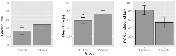
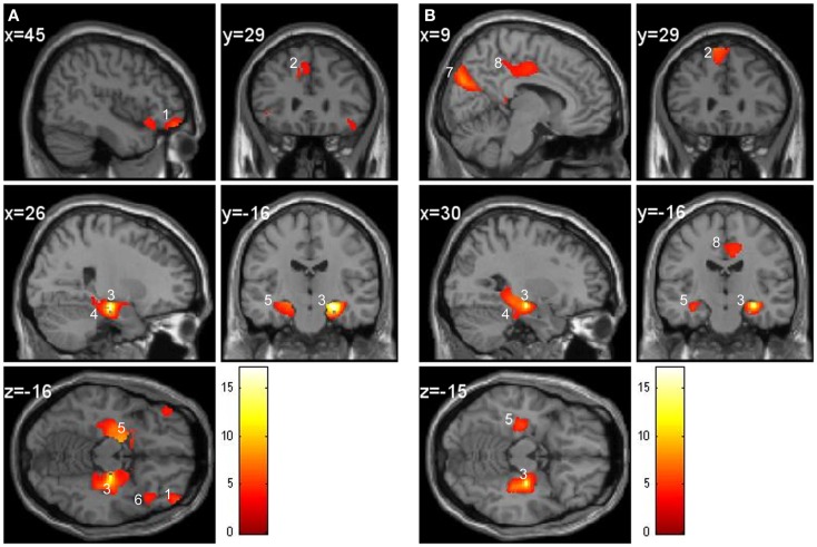
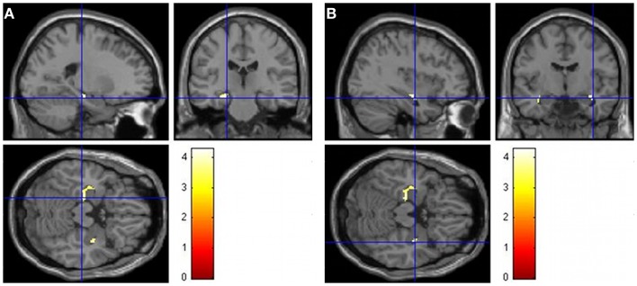
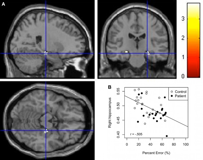
Episodic memory, related to the hippocampus, has been found to be impaired in schizophrenia. Further, hippocampal anomalies have also been observed in schizophrenia. This study investigated whether average hippocampal gray matter (GM) would differentiate performance on a hippocampus-dependent memory task in patients with schizophrenia and healthy controls. Twenty-one patients with schizophrenia and 22 control participants were scanned with an MRI while being tested on a wayfinding task in a virtual town (e.g., find the grocery store from the school). Regressions were performed for both groups individually and together using GM and performance on the wayfinding task. Results indicate that controls successfully completed the task more often than patients, took less time, and made fewer errors. Additionally, controls had significantly more hippocampal GM than patients. Poor performance was associated with a GM decrease in the right hippocampus for both groups. Within group regressions found an association between right hippocampi GM and performance in controls and an association between the left hippocampi GM and performance in patients. A second analysis revealed that different anatomical GM regions, known to be associated with the hippocampus, such as the parahippocampal cortex, amygdala, medial, and orbital prefrontal cortices, covaried with the hippocampus in the control group. Interestingly, the cuneus and cingulate gyrus also covaried with the hippocampus in the patient group but the orbital frontal cortex did not, supporting the hypothesis of impaired connectivity between the hippocampus and the frontal cortex in schizophrenia. These results present important implications for creating intervention programs aimed at measuring functional and structural changes in the hippocampus in schizophrenia.
与海马体相关的情景记忆在精神分裂症患者中已被发现受损。此外,在精神分裂症患者中也观察到了海马体异常。本研究调查了平均海马体灰质(GM)是否能区分精神分裂症患者和健康对照者在依赖海马体的记忆任务中的表现。21名精神分裂症患者和22名对照参与者在接受磁共振成像扫描时,同时在一个虚拟城镇中进行寻路任务测试(例如,从学校找到杂货店)。分别对两组以及将两组合并使用GM和寻路任务表现进行回归分析。结果表明,对照组比患者更频繁地成功完成任务,花费时间更少,且犯错更少。此外,对照组的海马体GM明显多于患者。两组中表现不佳均与右侧海马体GM减少有关。组内回归分析发现,对照组右侧海马体GM与表现之间存在关联,而患者组左侧海马体GM与表现之间存在关联。第二项分析显示,在对照组中,已知与海马体相关的不同解剖学GM区域,如海马旁皮质、杏仁核、内侧和眶额皮质,与海马体共同变化。有趣的是,楔叶和扣带回在患者组中也与海马体共同变化,但眶额皮质没有,这支持了精神分裂症中海马体与额叶皮质之间连接受损的假说。这些结果对于创建旨在测量精神分裂症中海马体功能和结构变化的干预项目具有重要意义。