Piñar Guadalupe, Sterflinger Katja, Pinzari Flavia
Institute of Applied Microbiology, Department of Biotechnology, University of Natural Resources and Life Sciences, Muthgasse 11, Vienna, 1190, Austria.
Environ Microbiol. 2015 Feb;17(2):427-43. doi: 10.1111/1462-2920.12471. Epub 2014 Apr 28.
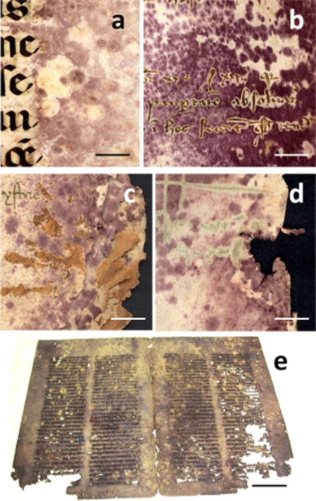
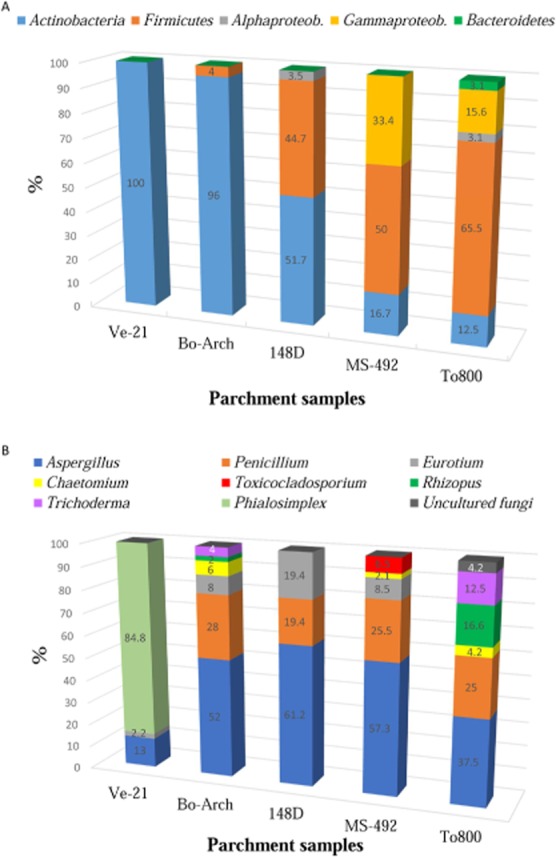
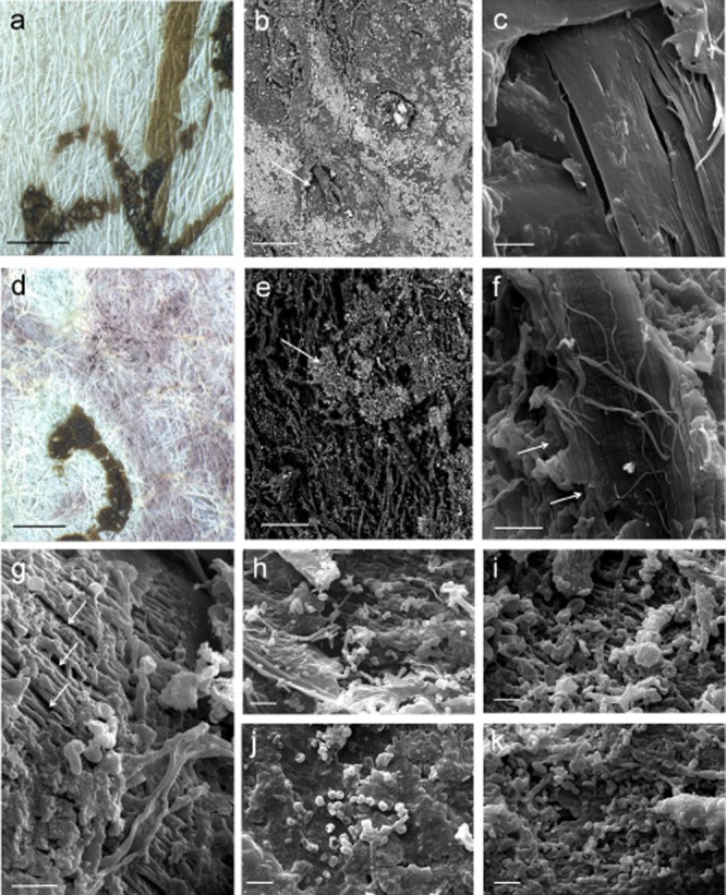
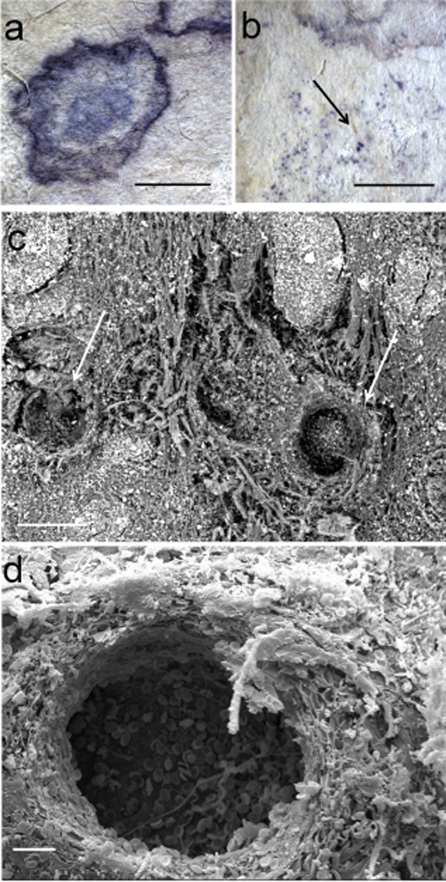
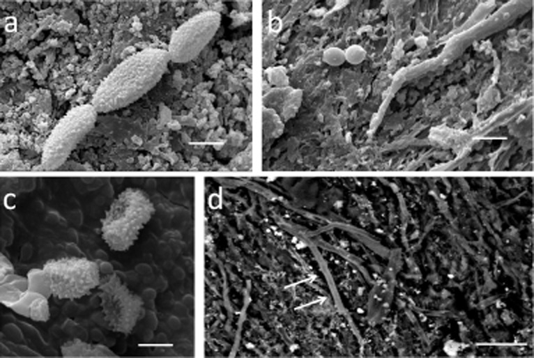
Many ancient parchments are defaced by red or purple maculae associated with localized destruction of collagen fibres. Although the main characteristics of this damage were present in most of the manuscripts analysed by many authors, no common microbial or fungal denominator has been found so far, and little or no correspondence between the microbial or fungal species isolated from materials could be addressed. In this study, culture-independent molecular methods and scanning electron microscopy (SEM) were used to identify fungal and bacterial communities on parchments affected by the purple stains. Protocols for c extraction and nucleic-acid-based strategies were selected for assays examining the community structure of fungi and bacteria on biodeteriorated parchment. Both SEM and molecular analysis detected the presence of bacterial and fungal cells in the damaged areas. Halophilic, halotolerant proteolytic bacterial species were selected by the saline environment provided by the parchment samples. As common microbial denominators, members of the Actinobacteria, mainly Saccharopolyspora spp. and species of Aspergillus, were detected in all investigated cases. It is proposed that a relationship exists between the phenomenon of purple spots on ancient parchments and that of the 'red heat' phenomenon, known to be present in some products manufactured with marine salt.
许多古代羊皮纸都被与胶原纤维局部破坏相关的红色或紫色斑点毁损。尽管许多作者分析的大多数手稿中都存在这种损伤的主要特征,但迄今为止尚未发现常见的微生物或真菌共性,而且从材料中分离出的微生物或真菌种类之间几乎没有对应关系。在本研究中,采用非培养分子方法和扫描电子显微镜(SEM)来鉴定受紫色污渍影响的羊皮纸上的真菌和细菌群落。选择了用于DNA提取的方案和基于核酸的策略来检测生物劣化羊皮纸上真菌和细菌的群落结构。SEM和分子分析均检测到受损区域存在细菌和真菌细胞。嗜盐、耐盐的蛋白水解细菌种类是由羊皮纸样本提供的盐环境选择出来的。作为常见的微生物共性,在所有调查案例中均检测到放线菌成员,主要是糖多孢菌属物种和曲霉属物种。有人提出,古代羊皮纸上的紫色斑点现象与某些用海盐制造的产品中存在的“红热”现象之间存在关联。