• 文献检索
  • 文档翻译
  • 深度研究
  • 学术资讯
  • Suppr Zotero 插件Zotero 插件
  • 邀请有礼
  • 套餐&价格
  • 历史记录
应用&插件
Suppr Zotero 插件Zotero 插件浏览器插件Mac 客户端Windows 客户端微信小程序
定价
高级版会员购买积分包购买API积分包
服务
文献检索文档翻译深度研究API 文档MCP 服务
关于我们
关于 Suppr公司介绍联系我们用户协议隐私条款
关注我们

Suppr 超能文献

核心技术专利:CN118964589B侵权必究
粤ICP备2023148730 号-1Suppr @ 2026

文献检索

告别复杂PubMed语法,用中文像聊天一样搜索,搜遍4000万医学文献。AI智能推荐,让科研检索更轻松。

立即免费搜索

文件翻译

保留排版,准确专业,支持PDF/Word/PPT等文件格式,支持 12+语言互译。

免费翻译文档

深度研究

AI帮你快速写综述,25分钟生成高质量综述,智能提取关键信息,辅助科研写作。

立即免费体验

用于微组织阵列扫描电化学显微镜分析的微冲压培养皿。

Microstamped Petri dishes for scanning electrochemical microscopy analysis of arrays of microtissues.

作者信息

Sridhar Adithya, de Boer Hans L, van den Berg Albert, Le Gac Séverine

机构信息

BIOS Lab on a Chip Group, MESA+ Institute of Nanotechnology, University of Twente, Enschede, The Netherlands.

出版信息

PLoS One. 2014 Apr 1;9(4):e93618. doi: 10.1371/journal.pone.0093618. eCollection 2014.

DOI:10.1371/journal.pone.0093618
PMID:24690887
原文链接:https://pmc.ncbi.nlm.nih.gov/articles/PMC3972177/
Abstract

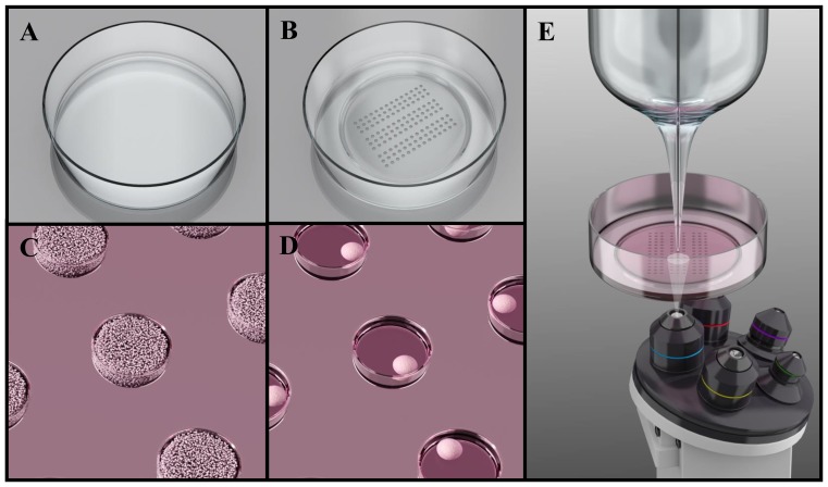
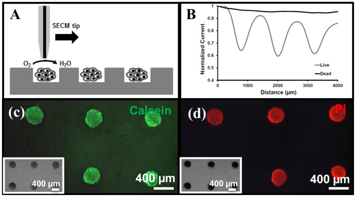
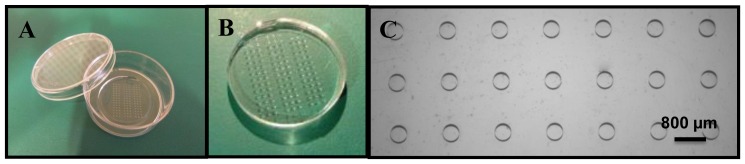
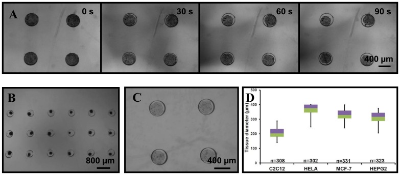
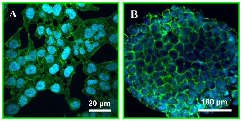
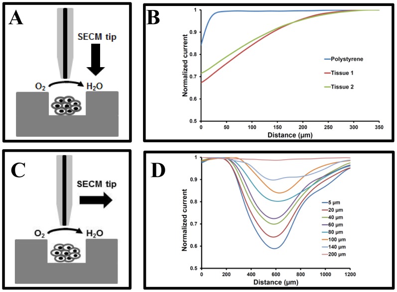
While scanning electrochemical microscopy (SECM) is a powerful technique for non-invasive analysis of cells, SECM-based assays remain scarce and have been mainly limited so far to single cells, which is mostly due to the absence of suitable platform for experimentation on 3D cellular aggregates or microtissues. Here, we report stamping of a Petri dish with a microwell array for large-scale production of microtissues followed by their in situ analysis using SECM. The platform is realized by hot embossing arrays of microwells (200 μm depth; 400 μm diameter) in commercially available Petri dishes, using a PDMS stamp. Microtissues form spontaneously in the microwells, which is demonstrated here using various cell lines (e.g., HeLa, C2C12, HepG2 and MCF-7). Next, the respiratory activity of live HeLa microtissues is assessed by monitoring the oxygen reduction current in constant height mode and at various distances above the platform surface. Typically, at a 40 μm distance from the microtissue, a 30% decrease in the oxygen reduction current is measured, while above 250 μm, no influence of the presence of the microtissues is detected. After exposure to a model drug (50% ethanol), no such changes in oxygen concentration are found at any height in solution, which reflects that microtissues are not viable anymore. This is furthermore confirmed using conventional live/dead fluorescent stains. This live/dead assay demonstrates the capability of the proposed approach combining SECM and microtissue arrays formed in a stamped Petri dish for conducting cellular assays in a non-invasive way on 3D cellular models.

摘要

扫描电化学显微镜(SECM)是一种用于细胞非侵入性分析的强大技术,但基于SECM的检测方法仍然很少,到目前为止主要局限于单细胞检测,这主要是由于缺乏适用于三维细胞聚集体或微组织实验的合适平台。在此,我们报道了一种在培养皿上冲压微孔阵列以大规模生产微组织,然后使用SECM对其进行原位分析的方法。该平台是通过使用聚二甲基硅氧烷(PDMS)印章在市售培养皿上热压印微孔阵列(深度200μm;直径400μm)来实现的。微组织在微孔中自发形成,这里使用多种细胞系(例如,HeLa、C2C12、HepG2和MCF-7)进行了证明。接下来,通过在恒定高度模式下监测平台表面上方不同距离处的氧还原电流,评估活HeLa微组织的呼吸活性。通常,在距微组织40μm的距离处,测量到氧还原电流降低了30%,而在250μm以上,未检测到微组织存在的影响。在暴露于模型药物(50%乙醇)后,在溶液中的任何高度都未发现氧浓度有此类变化,这表明微组织不再具有活力。使用传统的活/死荧光染色进一步证实了这一点。这种活/死检测证明了所提出的方法结合SECM和在冲压培养皿中形成的微组织阵列以对三维细胞模型进行非侵入性细胞检测的能力。

https://cdn.ncbi.nlm.nih.gov/pmc/blobs/5ab3/3972177/9a982b37be76/pone.0093618.g006.jpg
https://cdn.ncbi.nlm.nih.gov/pmc/blobs/5ab3/3972177/3929b0208cea/pone.0093618.g001.jpg
https://cdn.ncbi.nlm.nih.gov/pmc/blobs/5ab3/3972177/0c00d305c048/pone.0093618.g002.jpg
https://cdn.ncbi.nlm.nih.gov/pmc/blobs/5ab3/3972177/5ce06f0b6acb/pone.0093618.g003.jpg
https://cdn.ncbi.nlm.nih.gov/pmc/blobs/5ab3/3972177/8d9b0392a2fe/pone.0093618.g004.jpg
https://cdn.ncbi.nlm.nih.gov/pmc/blobs/5ab3/3972177/1b2f204ae545/pone.0093618.g005.jpg
https://cdn.ncbi.nlm.nih.gov/pmc/blobs/5ab3/3972177/9a982b37be76/pone.0093618.g006.jpg

相似文献

1
Microstamped Petri dishes for scanning electrochemical microscopy analysis of arrays of microtissues.用于微组织阵列扫描电化学显微镜分析的微冲压培养皿。
PLoS One. 2014 Apr 1;9(4):e93618. doi: 10.1371/journal.pone.0093618. eCollection 2014.
2
Monitoring the cellular activity of a cultured single cell by scanning electrochemical microscopy (SECM). A comparison with fluorescence viability monitoring.通过扫描电化学显微镜(SECM)监测培养的单细胞的细胞活性。与荧光活力监测的比较。
Biosens Bioelectron. 2003 Oct 1;18(11):1379-83. doi: 10.1016/s0956-5663(03)00083-6.
3
Systematic Analysis of Different Cell Spheroids with a Microfluidic Device Using Scanning Electrochemical Microscopy and Gene Expression Profiling.利用扫描电化学显微镜和基因表达谱对微流控装置中不同细胞球的系统分析。
Anal Chem. 2019 Apr 2;91(7):4307-4311. doi: 10.1021/acs.analchem.9b00376. Epub 2019 Mar 15.
4
Large-scale cultivation of transplantable dermal papilla cellular aggregates using microfabricated PDMS arrays.使用微加工 PDMS 阵列大规模培养可移植的真皮乳头细胞聚集体。
Acta Biomater. 2011 Jan;7(1):315-24. doi: 10.1016/j.actbio.2010.08.012. Epub 2010 Aug 20.
5
Analysis of beat fluctuations and oxygen consumption in cardiomyocytes by scanning electrochemical microscopy.利用扫描电化学显微镜分析心肌细胞的搏动波动和耗氧量。
Anal Biochem. 2014 Feb 15;447:39-42. doi: 10.1016/j.ab.2013.11.008. Epub 2013 Nov 16.
6
The microwell-mesh: A novel device and protocol for the high throughput manufacturing of cartilage microtissues.微井网:一种用于高通量制造软骨微组织的新型装置和方案。
Biomaterials. 2015 Sep;62:1-12. doi: 10.1016/j.biomaterials.2015.05.013. Epub 2015 May 20.
7
Mapping Cd²⁺-induced membrane permeability changes of single live cells by means of scanning electrochemical microscopy.通过扫描电化学显微镜绘制镉离子诱导的单个活细胞的膜通透性变化图。
Anal Chim Acta. 2016 Feb 18;908:85-94. doi: 10.1016/j.aca.2015.12.027. Epub 2016 Jan 5.
8
Using the Microwell-mesh to culture microtissues in vitro and as a carrier to implant microtissues in vivo into mice.采用微井网格在体外培养微组织,并作为载体将微组织植入体内的小鼠中。
Sci Rep. 2021 Mar 4;11(1):5118. doi: 10.1038/s41598-021-84154-4.
9
High-Content Label-Free Single-Cell Analysis with a Microfluidic Device Using Programmable Scanning Electrochemical Microscopy.基于可编程扫描电化学显微镜的微流控芯片无标记高通量单细胞分析。
Anal Chem. 2021 Sep 14;93(36):12417-12425. doi: 10.1021/acs.analchem.1c02507. Epub 2021 Aug 31.
10
Analysing single live cells by scanning electrochemical microscopy.通过扫描电化学显微镜分析单个活细胞。
Analyst. 2019 Jan 28;144(3):738-752. doi: 10.1039/c8an01490f.

引用本文的文献

1
Extended Spherical Diffusion Theory: Electrochemiluminescence Imaging Analysis of Diffusive Molecules from Spherical Biosamples.扩展球形扩散理论:来自球形生物样本的扩散分子的电化学发光成像分析
Anal Chem. 2024 Dec 3;96(48):18967-18976. doi: 10.1021/acs.analchem.4c03167. Epub 2024 Nov 18.
2
Microarray-Based Electrochemical Biosensing.基于微阵列的电化学生物传感。
Adv Biochem Eng Biotechnol. 2024;187:317-338. doi: 10.1007/10_2023_229.
3
Evaluation of paclitaxel-loaded polymeric nanoparticles in 3D tumor model: impact of tumor stroma on penetration and efficacy.

本文引用的文献

1
Evaluation of the differentiation status of single embryonic stem cells using scanning electrochemical microscopy.利用扫描电化学显微镜评价单个胚胎干细胞的分化状态。
Chem Commun (Camb). 2013 Jul 25;49(58):6498-500. doi: 10.1039/c3cc43126f. Epub 2013 Jun 12.
2
Assessment of multidrug resistance on cell coculture patterns using scanning electrochemical microscopy.采用扫描电化学显微镜评估细胞共培养模式下的多药耐药性。
Proc Natl Acad Sci U S A. 2013 Jun 4;110(23):9249-54. doi: 10.1073/pnas.1214809110. Epub 2013 May 17.
3
Recent advances in high resolution scanning electrochemical microscopy of living cells--a review.
载紫杉醇聚合物纳米粒在 3D 肿瘤模型中的评价:肿瘤基质对渗透和疗效的影响。
Drug Deliv Transl Res. 2023 May;13(5):1470-1483. doi: 10.1007/s13346-023-01310-1. Epub 2023 Feb 28.
4
Three-Dimensional Printed Stamps for the Fabrication of Patterned Microwells and High-Throughput Production of Homogeneous Cell Spheroids.用于制造图案化微孔和高通量生产均匀细胞球体的三维打印印章
3D Print Addit Manuf. 2020 Jun 1;7(3):139-147. doi: 10.1089/3dp.2019.0129. Epub 2020 Jun 5.
5
Electrochemical imaging of cells and tissues.细胞和组织的电化学成像
Chem Sci. 2018 Apr 9;9(20):4546-4554. doi: 10.1039/c8sc01035h. eCollection 2018 May 28.
6
Single Cell Oxygen Mapping (SCOM) by Scanning Electrochemical Microscopy Uncovers Heterogeneous Intracellular Oxygen Consumption.单细胞氧测绘(SCOM)通过扫描电化学显微镜揭示了异质的细胞内氧消耗。
Sci Rep. 2017 Sep 12;7(1):11428. doi: 10.1038/s41598-017-11956-w.
7
Isolated Reporter Bacteria in Supramolecular Hydrogel Microwell Arrays.超分子水凝胶微井阵列中的分离报告细菌。
Langmuir. 2017 Aug 8;33(31):7799-7809. doi: 10.1021/acs.langmuir.7b00749. Epub 2017 May 31.
8
In vitro methods to study bubble-cell interactions: Fundamentals and therapeutic applications.研究气泡与细胞相互作用的体外方法:基础与治疗应用
Biomicrofluidics. 2016 Jan 28;10(1):011501. doi: 10.1063/1.4940429. eCollection 2016 Jan.
活细胞高分辨率扫描电化学显微镜的最新进展——综述。
Anal Chim Acta. 2013 May 2;775:1-13. doi: 10.1016/j.aca.2012.12.042. Epub 2013 Jan 4.
4
Local redox-cycling-based electrochemical chip device with deep microwells for evaluation of embryoid bodies.用于评估胚状体的带有深微孔的基于局部氧化还原循环的电化学芯片装置。
Angew Chem Int Ed Engl. 2012 Jul 2;51(27):6648-52. doi: 10.1002/anie.201201602. Epub 2012 May 25.
5
Opportunities and challenges for use of tumor spheroids as models to test drug delivery and efficacy.肿瘤球体作为药物输送和疗效测试模型的应用机会和挑战。
J Control Release. 2012 Dec 10;164(2):192-204. doi: 10.1016/j.jconrel.2012.04.045. Epub 2012 May 18.
6
A Pt layer/Pt disk electrode configuration to evaluate respiration and alkaline phosphatase activities of mouse embryoid bodies.一种 Pt 层/Pt 圆盘电极构型,用于评估小鼠胚体的呼吸和碱性磷酸酶活性。
Talanta. 2012 May 30;94:30-5. doi: 10.1016/j.talanta.2012.01.059. Epub 2012 Mar 3.
7
Tissue deformation spatially modulates VEGF signaling and angiogenesis.组织变形在空间上调节 VEGF 信号转导和血管生成。
Proc Natl Acad Sci U S A. 2012 May 1;109(18):6886-91. doi: 10.1073/pnas.1201626109. Epub 2012 Apr 17.
8
Engineers are from PDMS-land, Biologists are from Polystyrenia.工程师来自 PDMS 领域,生物学家来自聚苯乙烯领域。
Lab Chip. 2012 Apr 7;12(7):1224-37. doi: 10.1039/c2lc20982a. Epub 2012 Feb 8.
9
Monitoring oxygen consumption of single mouse embryos using an integrated electrochemical microdevice.利用集成电化学微器件监测单个小鼠胚胎的耗氧量。
Biosens Bioelectron. 2011 Dec 15;30(1):100-6. doi: 10.1016/j.bios.2011.08.037. Epub 2011 Sep 6.
10
Benchtop micromolding of polystyrene by soft lithography.通过软光刻技术进行聚苯乙烯的台式微成型。
Lab Chip. 2011 Sep 21;11(18):3089-97. doi: 10.1039/c1lc20281b. Epub 2011 Aug 2.