Institute for the Treatment of Esophageal and Thoracic Disease, Allegheny Health Network, Pittsburgh, Pennsylvania, United States of America.
International Scholars Program, University of Pittsburgh Medical Center, Pittsburgh, Pennsylvania, United States of America.
PLoS One. 2014 Apr 4;9(4):e93694. doi: 10.1371/journal.pone.0093694. eCollection 2014.
To assess the reliability of magnetic resonance imaging (MRI) for detection of esophageal cancer in the Levrat model of end-to-side esophagojejunostomy.
The Levrat model has proven utility in terms of its ability to replicate Barrett's carcinogenesis by inducing gastroduodenoesophageal reflux (GDER). Due to lack of data on the utility of non-invasive methods for detection of esophageal cancer, treatment efficacy studies have been limited, as adenocarcinoma histology has only been validated post-mortem. It would therefore be of great value if the validity and reliability of MRI could be established in this setting.
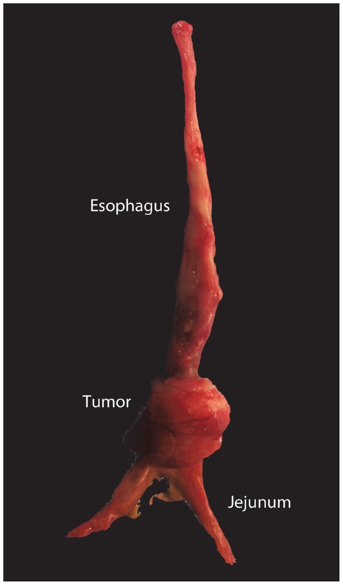
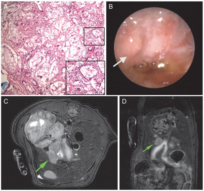
Chronic GDER reflux was induced in 19 male Sprague-Dawley rats using the modified Levrat model. At 40 weeks post-surgery, all animals underwent endoscopy, MRI scanning, and post-mortem histological analysis of the esophagus and anastomosis. With post-mortem histology serving as the gold standard, assessment of presence of esophageal cancer was made by five esophageal specialists and five radiologists on endoscopy and MRI, respectively.
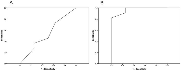
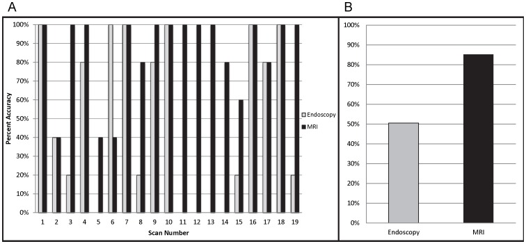
The accuracy of MRI and endoscopic analysis to correctly identify cancer vs. no cancer was 85.3% and 50.5%, respectively. ROC curves demonstrated that MRI rating had an AUC of 0.966 (p<0.001) and endoscopy rating had an AUC of 0.534 (p = 0.804). The sensitivity and specificity of MRI for identifying cancer vs. no-cancer was 89.1% and 80% respectively, as compared to 45.5% and 57.5% for endoscopy. False positive rates of MRI and endoscopy were 20% and 42.5%, respectively.
MRI is a more reliable diagnostic method than endoscopy in the Levrat model. The non-invasiveness of the tool and its potential to volumetrically quantify the size and number of tumors likely makes it even more useful in evaluating novel agents and their efficacy in treatment studies of esophageal cancer.
评估磁共振成像(MRI)在 Levrat 食管胃吻合术侧侧吻合模型中检测食管癌的可靠性。
Levrat 模型已被证明具有通过诱导胃食管反流(GDER)来复制 Barrett 癌变的能力。由于缺乏用于检测食管癌的非侵入性方法的效用数据,因此治疗效果研究受到限制,因为腺癌组织学仅在死后得到验证。因此,如果能够在此环境中确定 MRI 的有效性和可靠性,将具有非常重要的价值。
使用改良的 Levrat 模型在 19 只雄性 Sprague-Dawley 大鼠中诱导慢性 GDER 反流。手术后 40 周,所有动物均进行内镜检查、MRI 扫描以及食管和吻合口的死后组织学分析。以死后组织学为金标准,由五名食管专家和五名放射科医生分别在内镜和 MRI 上评估是否存在食管癌。
MRI 和内镜分析正确识别癌症与非癌症的准确性分别为 85.3%和 50.5%。ROC 曲线表明,MRI 评分的 AUC 为 0.966(p<0.001),内镜评分的 AUC 为 0.534(p=0.804)。MRI 用于识别癌症与非癌症的敏感性和特异性分别为 89.1%和 80%,而内镜分别为 45.5%和 57.5%。MRI 和内镜的假阳性率分别为 20%和 42.5%。
在 Levrat 模型中,MRI 是一种比内镜更可靠的诊断方法。该工具的非侵入性及其定量评估肿瘤大小和数量的潜力使其在评估新型药物及其在食管癌治疗研究中的疗效方面更加有用。