Division of Functional Neuroanatomy, Institute of Anatomy, University of Zürich Zürich, Switzerland.
Department of Zoology and Entomology, University of Pretoria Pretoria, South Africa.
Front Neuroanat. 2014 May 20;8:39. doi: 10.3389/fnana.2014.00039. eCollection 2014.
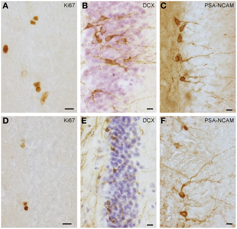
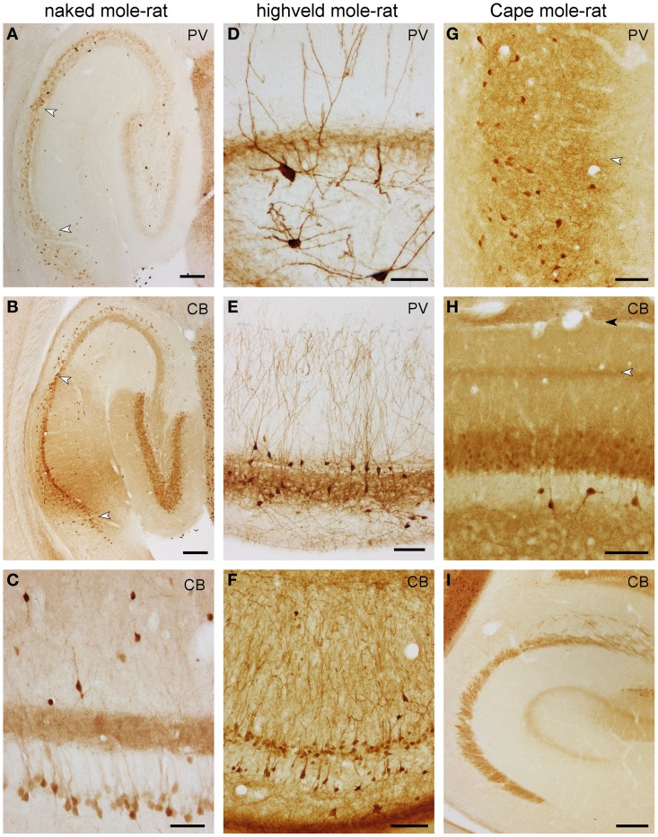
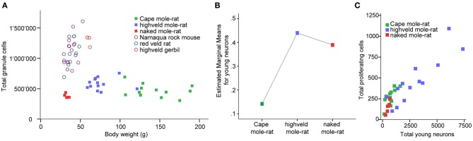
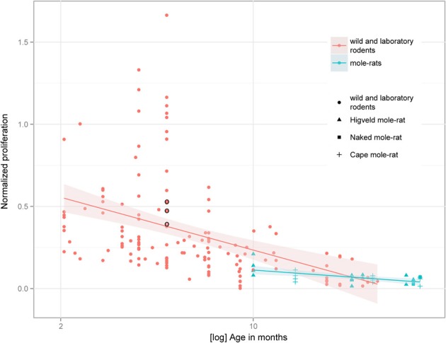
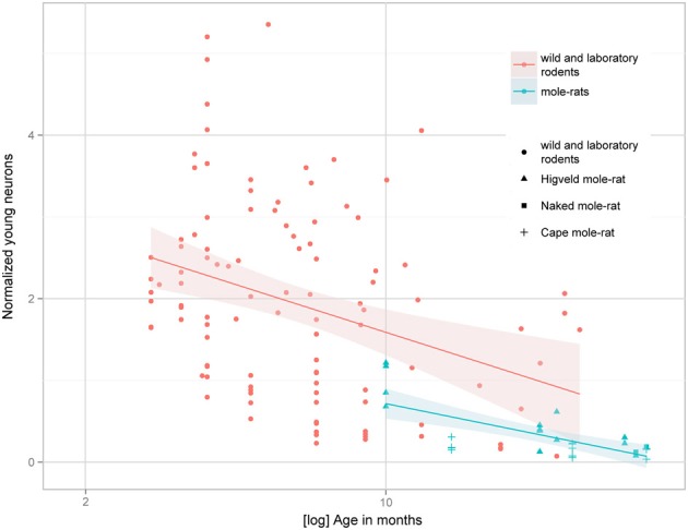
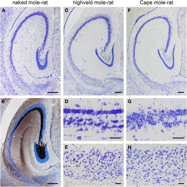
African mole-rats (family Bathyergidae) are small to medium sized, long-lived, and strictly subterranean rodents that became valuable animal models as a result of their longevity and diversity in social organization. The formation and integration of new hippocampal neurons in adult mammals (adult hippocampal neurogenesis, AHN) correlates negatively with age and positively with habitat complexity. Here we present quantitative data on AHN in wild-derived mole-rats of 1 year and older, and briefly describe its anatomical context including markers of neuronal function (calbindin and parvalbumin). Solitary Cape mole-rats (Georychus capensis), social highveld mole-rats (Cryptomys hottentotus pretoriae), and eusocial naked mole-rats (Heterocephalus glaber) were assessed. Compared to other rodents, the hippocampal formation in mole-rats is small, but shows a distinct cytoarchitecture in the dentate gyrus and CA1. Distributions of the calcium-binding proteins differ from those seen in rodents; e.g., calbindin in CA3 of naked mole-rats distributes similar to the pattern seen in early primate development, and calbindin staining extends into the stratum lacunosum-moleculare of Cape mole-rats. Proliferating cells and young neurons are found in low numbers in the hippocampus of all three mole-rat species. Resident granule cell numbers are low as well. Proliferating cells expressed as a percentage of resident granule cells are in the range of other rodents, while the percentage of young neurons is lower than that observed in surface dwelling rodents. Between mole-rat species, we observed no difference in the percentage of proliferating cells. The percentages of young neurons are high in social highveld and naked mole-rats, and low in solitary Cape mole-rats. The findings support that proliferation is regulated independently of average life expectancy and habitat. Instead, neuronal differentiation reflects species-specific demands, which appear lower in subterranean rodents.
非洲鼹鼠(Bathyergidae 科)是小型到中型、长寿命、严格的地下啮齿动物,由于其长寿和社会组织多样性,成为有价值的动物模型。成年哺乳动物(成年海马神经发生,AHN)中新海马神经元的形成和整合与年龄呈负相关,与栖息地复杂性呈正相关。本文提供了野生衍生的 1 岁及以上鼹鼠 AHN 的定量数据,并简要描述了其解剖学背景,包括神经元功能的标志物(钙结合蛋白和 parvalbumin)。独居海角鼹鼠(Georychus capensis)、社会性高地鼹鼠(Cryptomys hottentotus pretoriae)和社会性裸鼹鼠(Heterocephalus glaber)进行了评估。与其他啮齿动物相比,鼹鼠的海马结构较小,但在齿状回和 CA1 中具有明显的细胞结构。钙结合蛋白的分布与啮齿动物不同;例如,裸鼹鼠 CA3 中的钙结合蛋白分布类似于早期灵长类动物发育中所见的模式,钙结合蛋白染色延伸到海角鼹鼠的腔隙分子层。在所有三种鼹鼠物种的海马中,增殖细胞和年轻神经元的数量都很少。驻留颗粒细胞的数量也很低。作为驻留颗粒细胞的百分比表达的增殖细胞在其他啮齿动物的范围内,而年轻神经元的百分比低于在地面栖息的啮齿动物中观察到的百分比。在鼹鼠物种之间,我们观察到增殖细胞的百分比没有差异。年轻神经元的百分比在社会性高地和裸鼹鼠中较高,而在独居海角鼹鼠中较低。这些发现支持增殖是独立于平均预期寿命和栖息地而调节的。相反,神经元分化反映了物种特异性的需求,在地下啮齿动物中似乎较低。