• 文献检索
  • 文档翻译
  • 深度研究
  • 学术资讯
  • Suppr Zotero 插件Zotero 插件
  • 邀请有礼
  • 套餐&价格
  • 历史记录
应用&插件
Suppr Zotero 插件Zotero 插件浏览器插件Mac 客户端Windows 客户端微信小程序
定价
高级版会员购买积分包购买API积分包
服务
文献检索文档翻译深度研究API 文档MCP 服务
关于我们
关于 Suppr公司介绍联系我们用户协议隐私条款
关注我们

Suppr 超能文献

核心技术专利:CN118964589B侵权必究
粤ICP备2023148730 号-1Suppr @ 2026

文献检索

告别复杂PubMed语法,用中文像聊天一样搜索,搜遍4000万医学文献。AI智能推荐,让科研检索更轻松。

立即免费搜索

文件翻译

保留排版,准确专业,支持PDF/Word/PPT等文件格式,支持 12+语言互译。

免费翻译文档

深度研究

AI帮你快速写综述,25分钟生成高质量综述,智能提取关键信息,辅助科研写作。

立即免费体验

对稀疏特征进行皮质动力学编码。

Encoding cortical dynamics in sparse features.

机构信息

Athinoula A. Martinos Center for Biomedical Imaging, Massachusetts General Hospital/Harvard Medical School/Massachusetts Institute of Technology , Charlestown, MA , USA ; McGovern Institute, Massachusetts Institute of Technology , Cambridge, MA , USA ; Department of Neurology, Massachusetts General Hospital, Harvard Medical School , Boston, MA , USA.

Aix Marseille Université, CNRS, ENSAM, Université de Toulon, LSIS UMR 7296 , Marseille , France.

出版信息

Front Hum Neurosci. 2014 May 23;8:338. doi: 10.3389/fnhum.2014.00338. eCollection 2014.

DOI:10.3389/fnhum.2014.00338
PMID:24904377
原文链接:https://pmc.ncbi.nlm.nih.gov/articles/PMC4033054/
Abstract

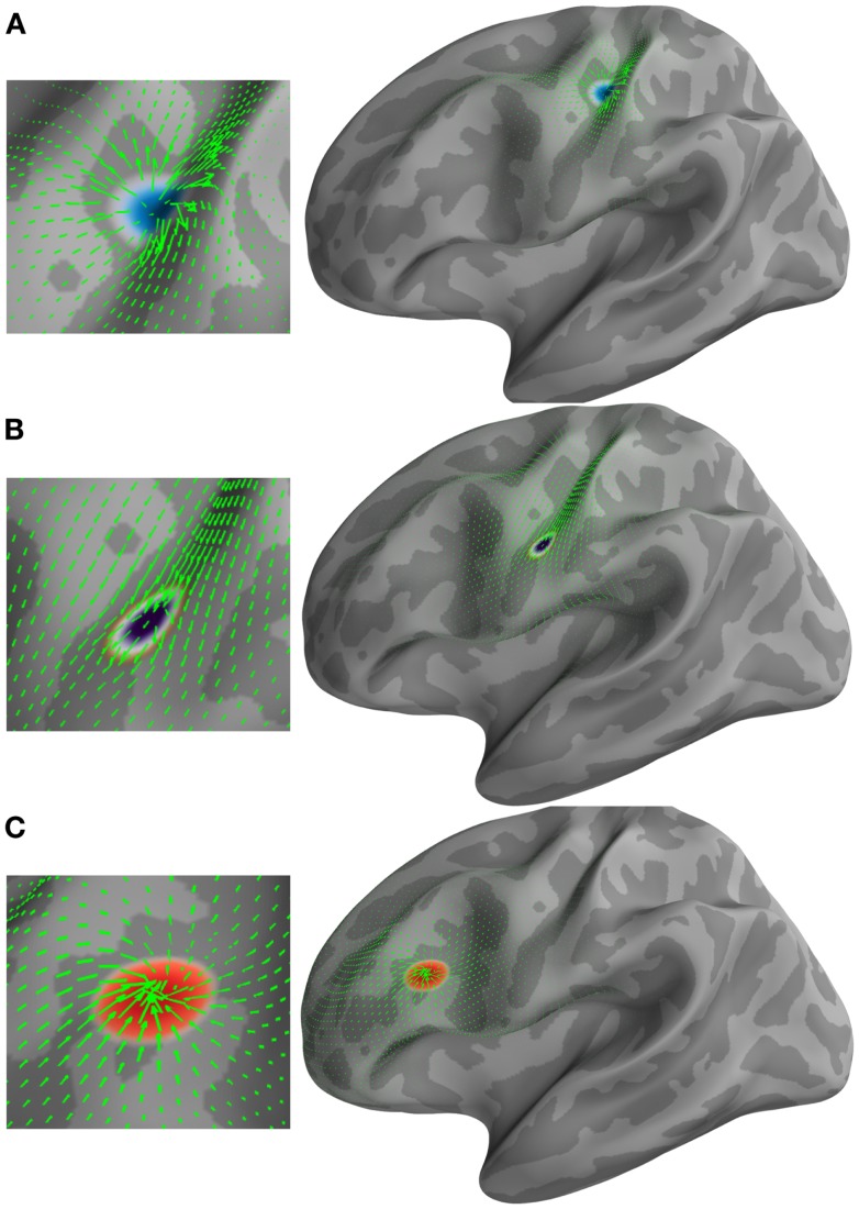
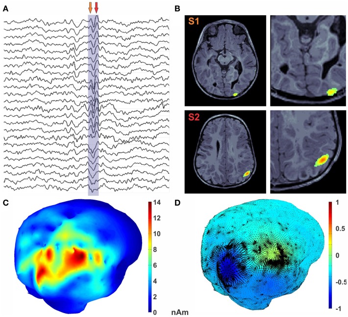
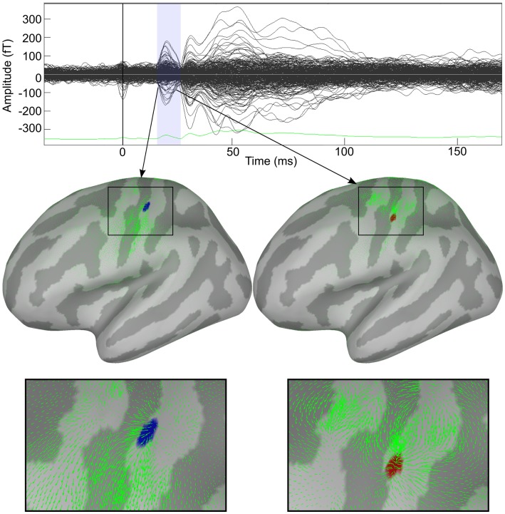
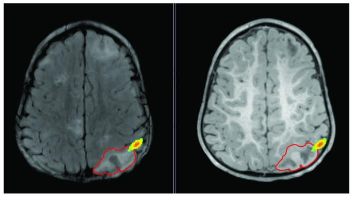
Distributed cortical solutions of magnetoencephalography (MEG) and electroencephalography (EEG) exhibit complex spatial and temporal dynamics. The extraction of patterns of interest and dynamic features from these cortical signals has so far relied on the expertise of investigators. There is a definite need in both clinical and neuroscience research for a method that will extract critical features from high-dimensional neuroimaging data in an automatic fashion. We have previously demonstrated the use of optical flow techniques for evaluating the kinematic properties of motion field projected on non-flat manifolds like in a cortical surface. We have further extended this framework to automatically detect features in the optical flow vector field by using the modified and extended 2-Riemannian Helmholtz-Hodge decomposition (HHD). Here, we applied these mathematical models on simulation and MEG data recorded from a healthy individual during a somatosensory experiment and an epilepsy pediatric patient during sleep. We tested whether our technique can automatically extract salient dynamical features of cortical activity. Simulation results indicated that we can precisely reproduce the simulated cortical dynamics with HHD; encode them in sparse features and represent the propagation of brain activity between distinct cortical areas. Using HHD, we decoded the somatosensory N20 component into two HHD features and represented the dynamics of brain activity as a traveling source between two primary somatosensory regions. In the epilepsy patient, we displayed the propagation of the epileptic activity around the margins of a brain lesion. Our findings indicate that HHD measures computed from cortical dynamics can: (i) quantitatively access the cortical dynamics in both healthy and disease brain in terms of sparse features and dynamic brain activity propagation between distinct cortical areas, and (ii) facilitate a reproducible, automated analysis of experimental and clinical MEG/EEG source imaging data.

摘要

脑磁图(MEG)和脑电图(EEG)的分布式皮质解译表现出复杂的时空动力学。从这些皮质信号中提取感兴趣的模式和动态特征,迄今为止一直依赖于研究人员的专业知识。在临床和神经科学研究中,都明确需要一种能够自动从高维神经影像学数据中提取关键特征的方法。我们之前已经证明了使用光流技术来评估投影到非平坦流形(如皮质表面)上的运动场的运动学特性。我们进一步扩展了这个框架,通过使用修改和扩展的 2-黎曼流形亥姆霍兹-霍奇分解(HHD)自动检测光流矢量场中的特征。在这里,我们将这些数学模型应用于模拟和 MEG 数据,这些数据是从一名健康个体在体感实验期间以及一名癫痫儿科患者在睡眠期间记录的。我们测试了我们的技术是否可以自动提取皮质活动的显著动态特征。模拟结果表明,我们可以使用 HHD 精确地再现模拟的皮质动力学;将其编码为稀疏特征,并表示不同皮质区域之间的大脑活动的传播。使用 HHD,我们将体感 N20 成分解码为两个 HHD 特征,并将大脑活动的动力学表示为两个主要体感区域之间的移动源。在癫痫患者中,我们显示了脑损伤边缘周围的癫痫活动的传播。我们的研究结果表明,从皮质动力学计算得出的 HHD 测量值可以:(i)以稀疏特征和不同皮质区域之间的动态大脑活动传播的形式,定量地评估健康和患病大脑的皮质动力学;(ii)促进对实验和临床 MEG/EEG 源成像数据的可重复、自动化分析。

https://cdn.ncbi.nlm.nih.gov/pmc/blobs/b017/4033054/9422cdc37a4d/fnhum-08-00338-g004.jpg
https://cdn.ncbi.nlm.nih.gov/pmc/blobs/b017/4033054/7de1489f53ba/fnhum-08-00338-g001.jpg
https://cdn.ncbi.nlm.nih.gov/pmc/blobs/b017/4033054/a9082fb09b3c/fnhum-08-00338-g002.jpg
https://cdn.ncbi.nlm.nih.gov/pmc/blobs/b017/4033054/cd2163641c20/fnhum-08-00338-g003.jpg
https://cdn.ncbi.nlm.nih.gov/pmc/blobs/b017/4033054/9422cdc37a4d/fnhum-08-00338-g004.jpg

相似文献

1
Encoding cortical dynamics in sparse features.对稀疏特征进行皮质动力学编码。
Front Hum Neurosci. 2014 May 23;8:338. doi: 10.3389/fnhum.2014.00338. eCollection 2014.
2
Characterization of interictal epileptiform discharges with time-resolved cortical current maps using the helmholtz-hodge decomposition.基于亥姆霍兹-霍奇分解的时分辨皮质电流图对发作间期癫痫样放电的特征描述。
Front Neurol. 2012 Oct 10;3:138. doi: 10.3389/fneur.2012.00138. eCollection 2012.
3
Recording human electrocorticographic (ECoG) signals for neuroscientific research and real-time functional cortical mapping.记录用于神经科学研究和实时功能性皮层图谱绘制的人类皮层脑电图(ECoG)信号。
J Vis Exp. 2012 Jun 26(64):3993. doi: 10.3791/3993.
4
Imaging brain activation streams from optical flow computation on 2-Riemannian manifolds.基于二维黎曼流形上光流计算的脑激活流成像。
Inf Process Med Imaging. 2007;20:470-81. doi: 10.1007/978-3-540-73273-0_39.
5
Complex patterns of spatially extended generators of epileptic activity: Comparison of source localization methods cMEM and 4-ExSo-MUSIC on high resolution EEG and MEG data.复杂的空间扩展癫痫活动发生器模式:高分辨率 EEG 和 MEG 数据上 cMEM 和 4-ExSo-MUSIC 源定位方法的比较。
Neuroimage. 2016 Dec;143:175-195. doi: 10.1016/j.neuroimage.2016.08.044. Epub 2016 Aug 22.
6
Neuro-current response functions: A unified approach to MEG source analysis under the continuous stimuli paradigm.神经电流响应函数:连续刺激范式下脑磁图源分析的统一方法。
Neuroimage. 2020 May 1;211:116528. doi: 10.1016/j.neuroimage.2020.116528. Epub 2020 Jan 13.
7
Investigation of EEG and MEG source imaging accuracy in reconstructing extended cortical sources.脑电图(EEG)和脑磁图(MEG)源成像在重建扩展皮质源方面的准确性研究。
Annu Int Conf IEEE Eng Med Biol Soc. 2011;2011:7013-6. doi: 10.1109/IEMBS.2011.6091773.
8
MEG-EEG Information Fusion and Electromagnetic Source Imaging: From Theory to Clinical Application in Epilepsy.脑磁图-脑电图信息融合与电磁源成像:从理论到癫痫的临床应用
Brain Topogr. 2015 Nov;28(6):785-812. doi: 10.1007/s10548-015-0437-3. Epub 2015 May 28.
9
Estimation of neural dynamics from MEG/EEG cortical current density maps: application to the reconstruction of large-scale cortical synchrony.从脑磁图/脑电图皮质电流密度图估计神经动力学:在大规模皮质同步重建中的应用。
IEEE Trans Biomed Eng. 2002 Sep;49(9):975-87. doi: 10.1109/TBME.2002.802013.
10
Simultaneous EEG and MEG source reconstruction in sparse electromagnetic source imaging.稀疏电磁源成像中的 EEG 和 MEG 源重建的同时进行。
Hum Brain Mapp. 2013 Apr;34(4):775-95. doi: 10.1002/hbm.21473. Epub 2011 Nov 18.

引用本文的文献

1
A Brief Introduction to Magnetoencephalography (MEG) and Its Clinical Applications.脑磁图(MEG)简介及其临床应用
Brain Sci. 2022 Jun 15;12(6):788. doi: 10.3390/brainsci12060788.
2
MEG Source Imaging and Group Analysis Using VBMEG.使用VBMEG进行脑磁图源成像与组分析。
Front Neurosci. 2019 Mar 22;13:241. doi: 10.3389/fnins.2019.00241. eCollection 2019.
3
Reorganization of the somatosensory cortex in hemiplegic cerebral palsy associated with impaired sensory tracts.偏瘫性脑瘫相关感觉通路受损患者体感皮层的重组。

本文引用的文献

1
Localization of the epileptogenic foci in tuberous sclerosis complex: a pediatric case report.结节性硬化症致痫灶的定位:1 例儿科病例报告。
Front Hum Neurosci. 2014 Mar 26;8:175. doi: 10.3389/fnhum.2014.00175. eCollection 2014.
2
MNE software for processing MEG and EEG data.MEG 和 EEG 数据处理的 MNE 软件。
Neuroimage. 2014 Feb 1;86:446-60. doi: 10.1016/j.neuroimage.2013.10.027. Epub 2013 Oct 24.
3
Improved Localizadon of Cortical Activity by Combining EEG and MEG with MRI Cortical Surface Reconstruction: A Linear Approach.
Neuroimage Clin. 2017 Oct 19;17:198-212. doi: 10.1016/j.nicl.2017.10.021. eCollection 2018.
4
Current and Emerging Potential of Magnetoencephalography in the Detection and Localization of High-Frequency Oscillations in Epilepsy.脑磁图在癫痫高频振荡检测与定位中的当前及新出现的潜力
Front Neurol. 2017 Jan 30;8:14. doi: 10.3389/fneur.2017.00014. eCollection 2017.
5
Editorial on emerging neuroimaging tools for studying normal and abnormal human brain development.关于用于研究正常和异常人类大脑发育的新兴神经成像工具的社论
Front Hum Neurosci. 2015 Mar 11;9:127. doi: 10.3389/fnhum.2015.00127. eCollection 2015.
通过将 EEG 和 MEG 与 MRI 皮质表面重建相结合来提高皮质活动的本地化:一种线性方法。
J Cogn Neurosci. 1993 Spring;5(2):162-76. doi: 10.1162/jocn.1993.5.2.162.
4
Can magnetoencephalography track the afferent information flow along white matter thalamo-cortical fibers?脑磁图能否追踪沿白质丘脑-皮层纤维的传入信息流?
Neuroimage. 2012 Apr 2;60(2):1092-105. doi: 10.1016/j.neuroimage.2012.01.054. Epub 2012 Jan 15.
5
Brainstorm: a user-friendly application for MEG/EEG analysis.头脑风暴:一款用于 MEG/EEG 分析的用户友好型应用程序。
Comput Intell Neurosci. 2011;2011:879716. doi: 10.1155/2011/879716. Epub 2011 Apr 13.
6
Tracking cortical activity from M/EEG using graph cuts with spatiotemporal constraints.使用具有时空约束的图割技术从 M/EEG 中跟踪皮质活动。
Neuroimage. 2011 Feb 1;54(3):1930-41. doi: 10.1016/j.neuroimage.2010.09.087. Epub 2010 Oct 7.
7
BA3b and BA1 activate in a serial fashion after median nerve stimulation: direct evidence from combining source analysis of evoked fields and cytoarchitectonic probabilistic maps.正中神经刺激后,BA3b 和 BA1 依次激活:来自诱发电场源分析与细胞构筑概率图谱结合的直接证据。
Neuroimage. 2011 Jan 1;54(1):60-73. doi: 10.1016/j.neuroimage.2010.07.054. Epub 2010 Aug 4.
8
Propagation of epileptic spikes reconstructed from spatiotemporal magnetoencephalographic and electroencephalographic source analysis.从时空磁共振和脑电图源分析重建的癫痫棘波传播。
Neuroimage. 2010 Mar;50(1):217-22. doi: 10.1016/j.neuroimage.2009.12.033. Epub 2009 Dec 16.
9
MEG's ability to localise accurately weak transient neural sources.脑磁图准确探测微弱瞬态神经源的能力。
Clin Neurophysiol. 2009 Nov;120(11):1958-1970. doi: 10.1016/j.clinph.2009.08.018. Epub 2009 Sep 25.
10
Optical flow approaches to the identification of brain dynamics.用于识别脑动力学的光流方法。
Hum Brain Mapp. 2009 Jun;30(6):1887-97. doi: 10.1002/hbm.20781.