Suzuki Junji, Kanemaru Kazunori, Ishii Kuniaki, Ohkura Masamichi, Okubo Yohei, Iino Masamitsu
Department of Pharmacology, Graduate School of Medicine, The University of Tokyo, 7-3-1 Bunkyo-ku, Tokyo 113-0033, Japan.
Department of Pharmacology, School of Medicine, Yamagata University, 2-2-2 Iida-nishi, Yamagata 990-9585, Japan.
Nat Commun. 2014 Jun 13;5:4153. doi: 10.1038/ncomms5153.
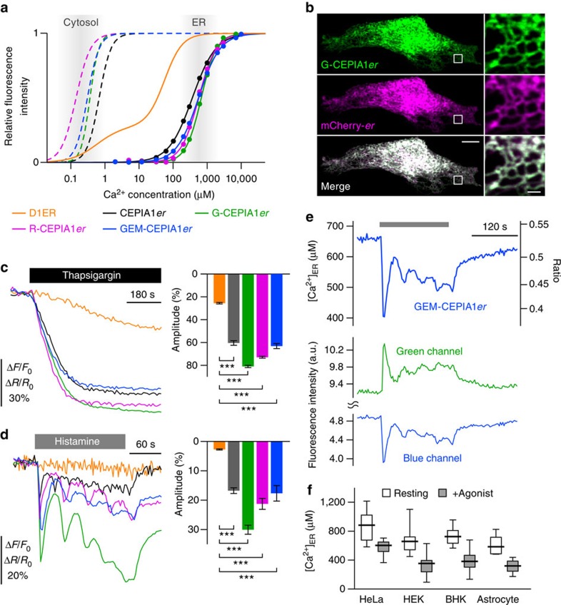
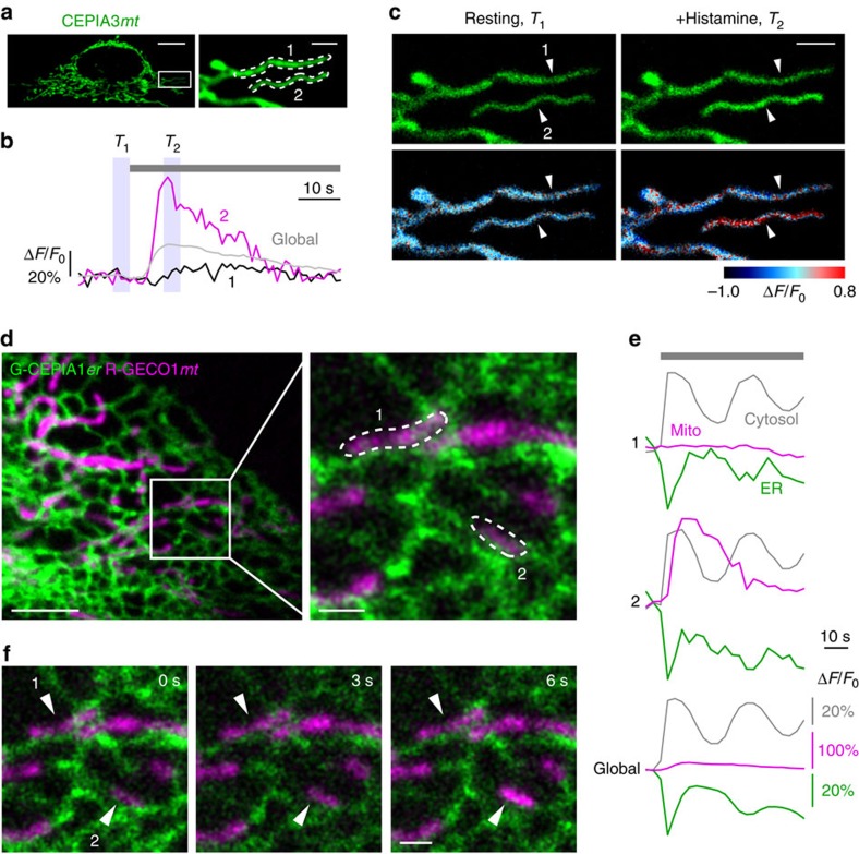
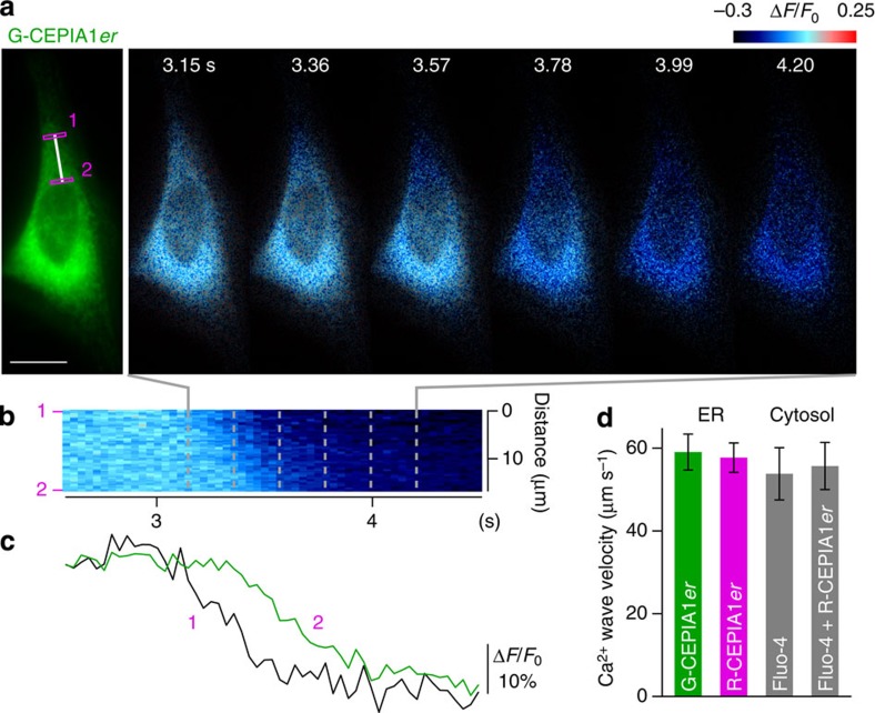
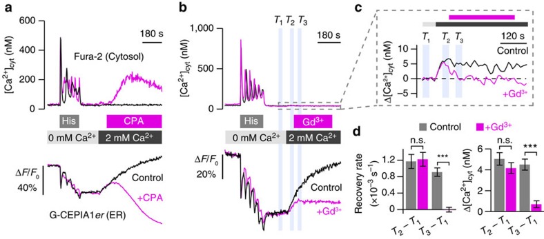
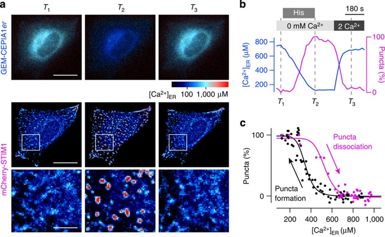
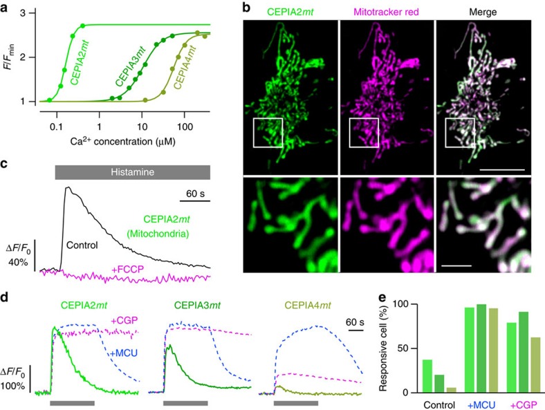
The endoplasmic reticulum (ER) and mitochondria accumulate Ca(2+) within their lumens to regulate numerous cell functions. However, determining the dynamics of intraorganellar Ca(2+) has proven to be difficult. Here we describe a family of genetically encoded Ca(2+) indicators, named calcium-measuring organelle-entrapped protein indicators (CEPIA), which can be utilized for intraorganellar Ca(2+) imaging. CEPIA, which emit green, red or blue/green fluorescence, are engineered to bind Ca(2+) at intraorganellar Ca(2+) concentrations. They can be targeted to different organelles and may be used alongside other fluorescent molecular markers, expanding the range of cell functions that can be simultaneously analysed. The spatiotemporal resolution of CEPIA makes it possible to resolve Ca(2+) import into individual mitochondria while simultaneously measuring ER and cytosolic Ca(2+). We have used these imaging capabilities to reveal differential Ca(2+) handling in individual mitochondria. CEPIA imaging is a useful new tool to further the understanding of organellar functions.
内质网(ER)和线粒体在其内腔中积累Ca(2+)以调节众多细胞功能。然而,确定细胞器内Ca(2+)的动态变化已被证明是困难的。在这里,我们描述了一类基因编码的Ca(2+)指示剂,名为细胞器捕获钙测量蛋白指示剂(CEPIA),其可用于细胞器内Ca(2+)成像。CEPIA可发出绿色、红色或蓝绿色荧光,经过设计可在细胞器内Ca(2+)浓度下结合Ca(2+)。它们可以靶向不同的细胞器,并可与其他荧光分子标记物一起使用,从而扩大了可同时分析的细胞功能范围。CEPIA的时空分辨率使得在同时测量内质网和细胞质Ca(2+)的情况下,能够分辨Ca(2+)进入单个线粒体的过程。我们利用这些成像能力揭示了单个线粒体中不同的Ca(2+)处理方式。CEPIA成像为进一步理解细胞器功能提供了一种有用的新工具。