Kim Moosang, Lee Seung-Jun
Department of Ophthalmology, School of Medicine, Kangwon National University, Chuncheon, Republic of Korea.
Clin Ophthalmol. 2014 May 29;8:1051-3. doi: 10.2147/OPTH.S63328. eCollection 2014.
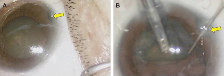
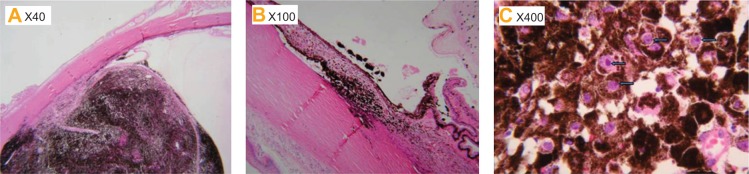
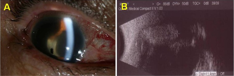
A 62-year-old female presented to our institution with dimness of vision in her right eye. On examination, her best corrected visual acuity was 20/100 in the right eye. The intraocular pressures were 14 mmHg in both eyes. Slit-lamp examination revealed nuclear sclerotic cataracts bilaterally and iridodialysis in her right eye. Seven days after the first visit, cataract surgery was performed without any complications. One year later, she presented to our institution with acute visual loss and ocular pain in the right eye. Best corrected visual acuity of the right eye was light perception and the intraocular pressure was 44 mmHg. Slit-lamp examination revealed a ciliary body mass with widespread pigment dispersion in the anterior segment. Due to no useful vision and uncontrolled pain, enucleation of the right eye was performed. Histopathologic examination revealed a melanocytoma of the ciliary body.
一名62岁女性因右眼视力模糊前来我院就诊。检查发现,其右眼最佳矫正视力为20/100。双眼眼压均为14 mmHg。裂隙灯检查显示双眼核性硬化性白内障,右眼虹膜根部离断。初诊7天后,患者接受了白内障手术,未出现任何并发症。一年后,她因右眼急性视力丧失和眼痛再次前来我院。右眼最佳矫正视力仅为光感,眼压为44 mmHg。裂隙灯检查发现睫状体肿物,前段广泛色素播散。由于视力无法恢复且疼痛无法控制,遂行右眼眼球摘除术。组织病理学检查显示为睫状体黑素细胞瘤。