Tsai Nga Wing, Ngai Chun Wai, Mok Ka Leung, Tsung James W
Adult Intensive Care Unit, Queen Mary Hospital, 102 Pok Fu Lam Road, Hong Kong, SAR, China.
Accident and Emergency Department, Ruttonjee Hospital, 266 Queen's Way, Wan Chai, Hong Kong, SAR, China.
Crit Ultrasound J. 2014 May 20;6(1):6. doi: 10.1186/2036-7902-6-6. eCollection 2014.
Lung ultrasound has been shown to identify in real-time, various pathologies of the lung such as pneumonia, viral pneumonia, and acute respiratory distress syndrome (ARDS). Lung ultrasound maybe a first-line alternative to chest X-ray and CT scan in critically ill patients with respiratory failure. We describe the use of lung ultrasound imaging and findings in two cases of severe respiratory failure from avian influenza A (H7N9) infection.
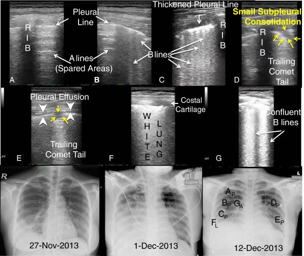
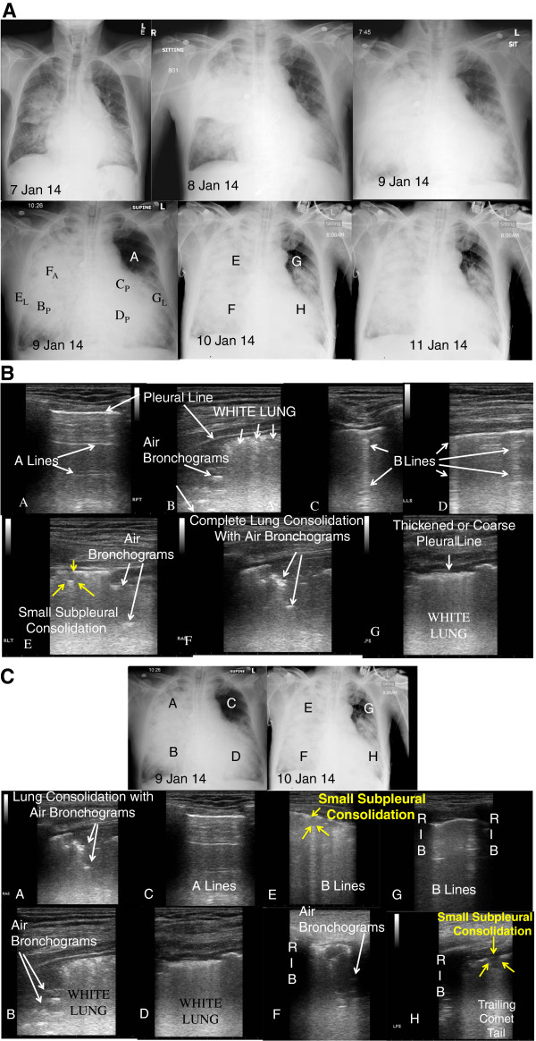
Serial lung ultrasound images and video from two cases of H7N9 respiratory failure requiring mechanical ventilation and extracorporeal membrane oxygenation in a tertiary care intensive care unit were analyzed for characteristic lung ultrasound findings described previously for respiratory failure and infection. These findings were followed serially, correlated with clinical course and chest X-ray.
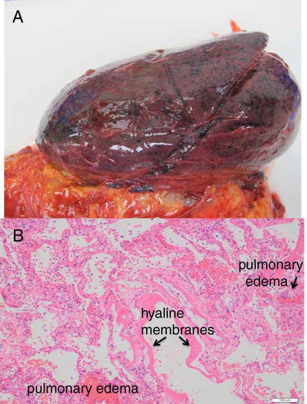
IN BOTH PATIENTS, CHARACTERISTIC LUNG ULTRASOUND FINDINGS HAVE BEEN OBSERVED AS PREVIOUSLY DESCRIBED IN VIRAL PULMONARY INFECTIONS: subpleural consolidations associated or not with local pleural effusion. In addition, numerous, confluent, or coalescing B-lines leading to 'white lung' with corresponding pleural line thickening are associated with ARDS. Extension or reduction of lesions observed with ultrasound was also correlated respectively with clinical worsening or improvement. Coexisting consolidated pneumonia with sonographic air bronchograms was noted in one patient who did not survive.
Clinicians with access to point-of-care ultrasonography may use these findings as an alternative to chest X-ray or CT scan. Lung ultrasound imaging may assist in the efficient allocation of intensive care for patients with respiratory failure from viral pulmonary infections, especially in resource scarce settings or situations such as future respiratory virus outbreaks or pandemics.
肺超声已被证明能够实时识别肺部的各种病变,如肺炎、病毒性肺炎和急性呼吸窘迫综合征(ARDS)。对于患有呼吸衰竭的重症患者,肺超声可能是胸部X线和CT扫描的一线替代检查方法。我们描述了在两例甲型H7N9禽流感感染导致严重呼吸衰竭患者中肺超声成像的应用及结果。
分析了在一家三级医疗重症监护病房中,两例因H7N9呼吸衰竭需要机械通气和体外膜肺氧合治疗的患者的系列肺超声图像和视频,以寻找先前描述的与呼吸衰竭和感染相关的特征性肺超声表现。对这些表现进行连续观察,并与临床病程和胸部X线检查结果进行关联分析。
在这两名患者中,均观察到了先前在病毒性肺部感染中描述过的特征性肺超声表现:胸膜下实变,可伴有或不伴有局部胸腔积液。此外,大量、融合或聚集的B线导致“白肺”并伴有相应的胸膜线增厚与ARDS相关。超声观察到的病变范围扩大或缩小也分别与临床病情恶化或改善相关。在一名死亡患者中发现了合并存在的实变肺炎及超声可见的空气支气管征。
能够进行床旁超声检查的临床医生可将这些表现作为胸部X线或CT扫描的替代方法。肺超声成像可为病毒性肺部感染导致呼吸衰竭的患者提供重症监护的有效分配依据,特别是在资源匮乏的环境中,或在未来呼吸道病毒爆发或大流行等情况下。