Chiu Chi-Li, Aguilar Jose S, Tsai Connie Y, Wu GuiKai, Gratton Enrico, Digman Michelle A
Department of Developmental and Cell Biology, University of California Irvine, Irvine, California, United States of America.
Department of Biomedical Engineering, Laboratory for Fluorescence Dynamics, University of California Irvine, Irvine, California, United States of America.
PLoS One. 2014 Jun 24;9(6):e99896. doi: 10.1371/journal.pone.0099896. eCollection 2014.
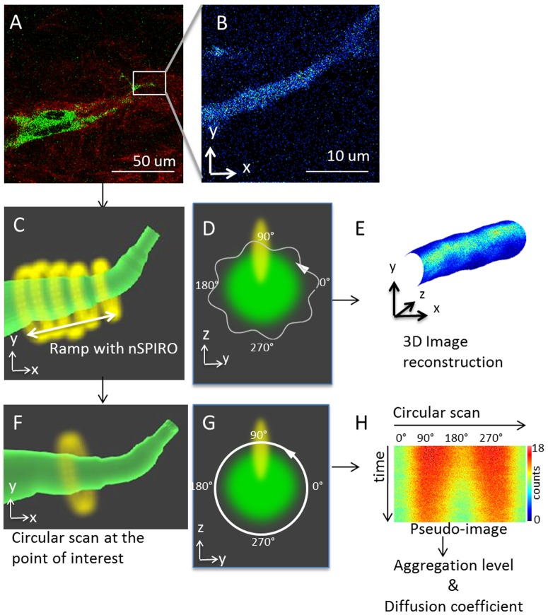
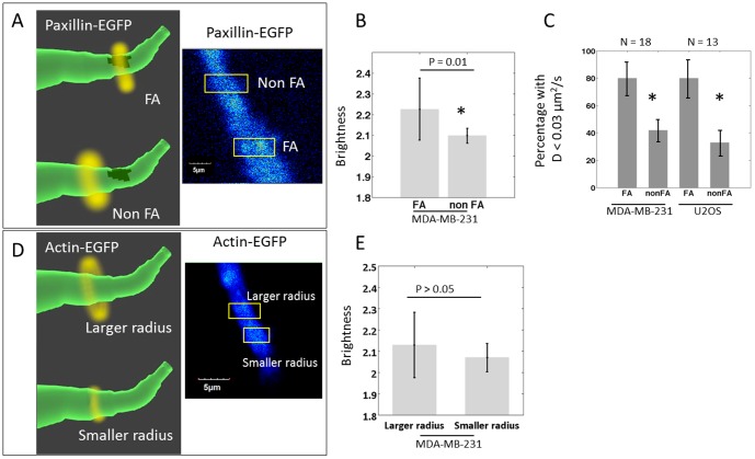
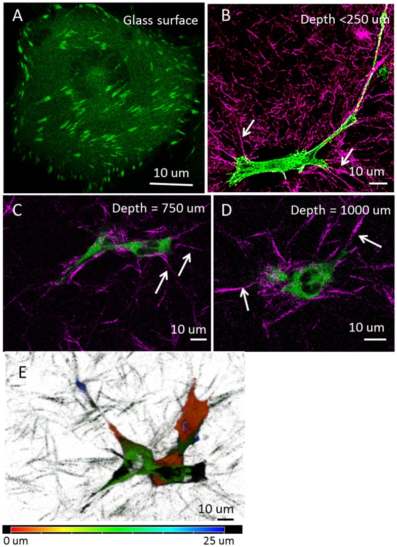
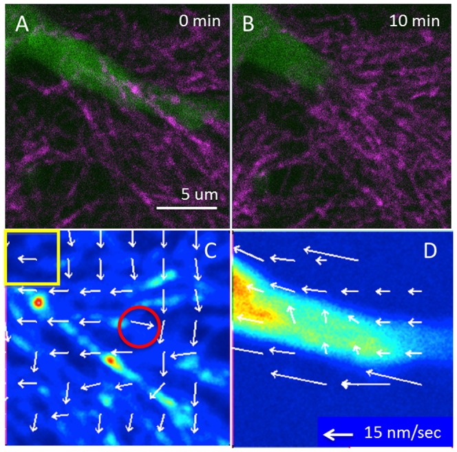
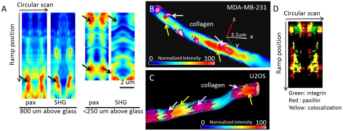
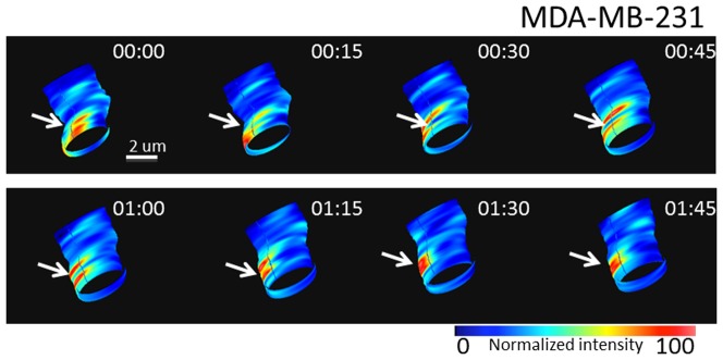
Organization and dynamics of focal adhesion proteins have been well characterized in cells grown on two-dimensional (2D) cell culture surfaces. However, much less is known about the dynamic association of these proteins in the 3D microenvironment. Limited imaging technologies capable of measuring protein interactions in real time and space for cells grown in 3D is a major impediment in understanding how proteins function under different environmental cues. In this study, we applied the nano-scale precise imaging by rapid beam oscillation (nSPIRO) technique and combined the scaning-fluorescence correlation spectroscopy (sFCS) and the number and molecular brightness (N&B) methods to investigate paxillin and actin dynamics at focal adhesions in 3D. Both MDA-MB-231 cells and U2OS cells produce elongated protrusions with high intensity regions of paxillin in cell grown in 3D collagen matrices. Using sFCS we found higher percentage of slow diffusing proteins at these focal spots, suggesting assembling/disassembling processes. In addition, the N&B analysis shows paxillin aggregated predominantly at these focal contacts which are next to collagen fibers. At those sites, actin showed slower apparent diffusion rate, which indicated that actin is either polymerizing or binding to the scaffolds in these locals. Our findings demonstrate that by multiplexing these techniques we have the ability to spatially and temporally quantify focal adhesion assembly and disassembly in 3D space and allow the understanding tumor cell invasion in a more complex relevant environment.
粘着斑蛋白的组织和动力学在二维(2D)细胞培养表面生长的细胞中已得到充分表征。然而,对于这些蛋白在三维微环境中的动态关联了解较少。能够实时测量三维培养细胞中蛋白相互作用的成像技术有限,这是理解蛋白在不同环境线索下如何发挥功能的主要障碍。在本研究中,我们应用了快速光束振荡纳米级精确成像(nSPIRO)技术,并结合扫描荧光相关光谱(sFCS)和数量与分子亮度(N&B)方法,来研究三维粘着斑处桩蛋白和肌动蛋白的动力学。在三维胶原基质中生长的MDA-MB-231细胞和U2OS细胞都会产生带有桩蛋白高强度区域的细长突起。使用sFCS,我们发现在这些粘着斑处慢扩散蛋白的比例更高,这表明存在组装/拆卸过程。此外,N&B分析显示桩蛋白主要聚集在靠近胶原纤维的这些粘着接触处。在这些位点,肌动蛋白显示出较慢的表观扩散速率,这表明肌动蛋白在这些局部区域要么正在聚合,要么正在与支架结合。我们的研究结果表明,通过复用这些技术,我们有能力在三维空间中对粘着斑的组装和拆卸进行时空定量,并有助于在更复杂的相关环境中理解肿瘤细胞的侵袭。