Jerman Urška Dragin, Kolenc Marko, Steyer Andrej, Veranič Peter, Prijatelj Mateja Poljšak, Kreft Mateja Erdani
Institute of Cell Biology, Faculty of Medicine, University of Ljubljana, SI-1000 Ljubljana, Slovenia.
Institute of Microbiology and Immunology, Faculty of Medicine, University of Ljubljana, SI-1000 Ljubljana, Slovenia.
Viruses. 2014 Jun 23;6(6):2505-18. doi: 10.3390/v6062505.
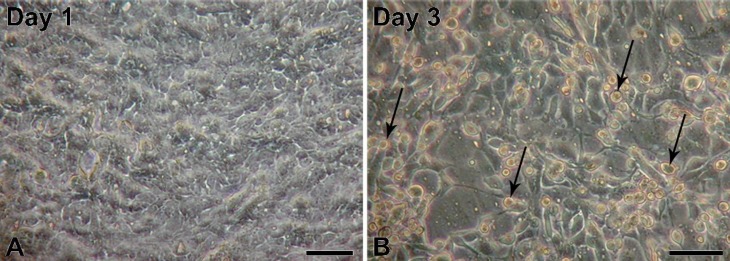
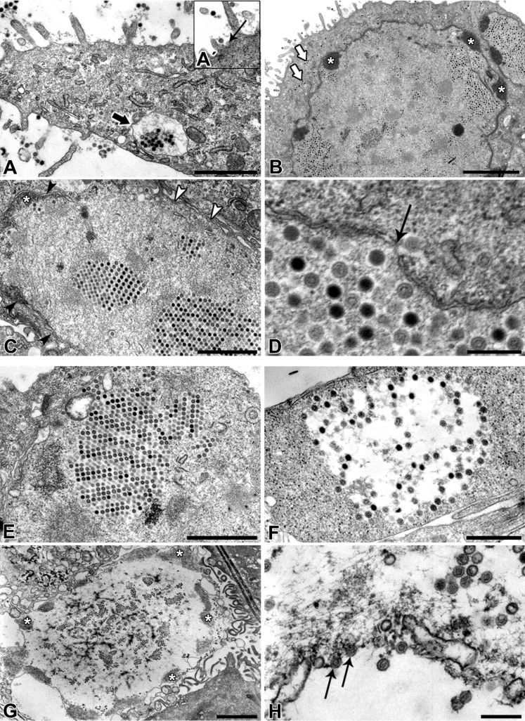
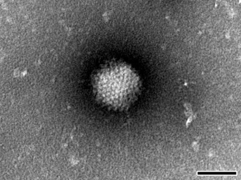
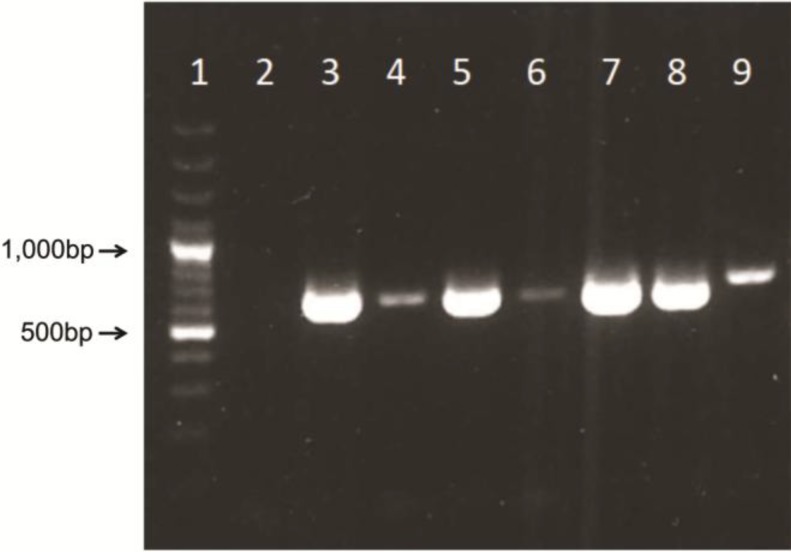
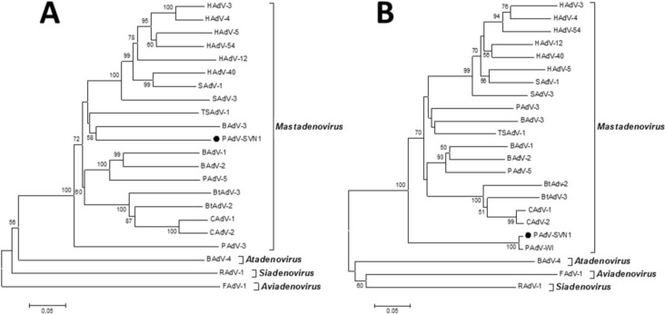
Contamination of cell cultures is the most common problem encountered in cell culture laboratories. Besides the secondary cell contaminations often occurring in the cell laboratories, the contaminations originating from donor animal or human tissue are equally as common, but usually harder to recognize and as such require special attention. The present study describes the detection of porcine adenovirus (PAdV), strain PAdV-SVN1 in cultures of normal porcine urothelial (NPU) cells isolated from urinary bladders of domestic pigs. NPU cell cultures were evaluated by light microscopy (LM), polymerase chain reaction (PCR), and additionally assessed by transmission electron microscopy (TEM). Characteristic ultrastructure of virions revealed the infection with adenovirus. The adenoviral contamination was further identified by the sequence analysis, which showed the highest similarity to recently described PAdV strain PAdV-WI. Additionally, the cell ultrastructural analysis confirmed the life-cycle characteristic for adenoviruses. To closely mimic the in vivo situation, the majority of research on in vitro models uses cell cultures isolated from human or animal tissue and their subsequent passages. Since the donor tissue could be a potential source of contamination, the microbiological screening of the excised tissue and harvested cell cultures is highly recommended.
细胞培养污染是细胞培养实验室中最常见的问题。除了细胞实验室中经常发生的二次细胞污染外,源自供体动物或人类组织的污染同样常见,但通常更难识别,因此需要特别关注。本研究描述了在家猪膀胱分离的正常猪尿路上皮(NPU)细胞培养物中检测猪腺病毒(PAdV),毒株PAdV-SVN1的情况。通过光学显微镜(LM)、聚合酶链反应(PCR)对NPU细胞培养物进行评估,并另外通过透射电子显微镜(TEM)进行评估。病毒粒子的特征超微结构显示存在腺病毒感染。通过序列分析进一步鉴定了腺病毒污染,结果表明其与最近描述的PAdV毒株PAdV-WI具有最高的相似性。此外,细胞超微结构分析证实了腺病毒的生命周期特征。为了紧密模拟体内情况,大多数体外模型研究使用从人类或动物组织分离的细胞培养物及其后续传代培养。由于供体组织可能是污染的潜在来源,强烈建议对切除的组织和收获的细胞培养物进行微生物学筛查。