• 文献检索
  • 文档翻译
  • 深度研究
  • 学术资讯
  • Suppr Zotero 插件Zotero 插件
  • 邀请有礼
  • 套餐&价格
  • 历史记录
应用&插件
Suppr Zotero 插件Zotero 插件浏览器插件Mac 客户端Windows 客户端微信小程序
定价
高级版会员购买积分包购买API积分包
服务
文献检索文档翻译深度研究API 文档MCP 服务
关于我们
关于 Suppr公司介绍联系我们用户协议隐私条款
关注我们

Suppr 超能文献

核心技术专利:CN118964589B侵权必究
粤ICP备2023148730 号-1Suppr @ 2026

文献检索

告别复杂PubMed语法,用中文像聊天一样搜索,搜遍4000万医学文献。AI智能推荐,让科研检索更轻松。

立即免费搜索

文件翻译

保留排版,准确专业,支持PDF/Word/PPT等文件格式,支持 12+语言互译。

免费翻译文档

深度研究

AI帮你快速写综述,25分钟生成高质量综述,智能提取关键信息,辅助科研写作。

立即免费体验

乳腺微钙化的光声成像:对8号针芯活检乳腺标本的初步研究

Photoacoustic imaging of breast microcalcifications: a preliminary study with 8-gauge core-biopsied breast specimens.

作者信息

Kim Ga Ram, Kang Jeeun, Kwak Jin Young, Chang Jin Ho, Kim Seung Il, Youk Ji Hyun, Moon Hee Jung, Kim Min Jung, Kim Eun-Kyung

机构信息

Department of Radiology and Research Institute of Radiological Science, Severance Hospital, Yonsei University College of Medicine, Seoul, Republic of Korea.

Sogang Institute of Advanced Technology, Sogang University, Seoul, Republic of Korea; Interdisciplinary Program of Integrated Biotechnology, Seoul, Republic of Korea.

出版信息

PLoS One. 2014 Aug 25;9(8):e105878. doi: 10.1371/journal.pone.0105878. eCollection 2014.

DOI:10.1371/journal.pone.0105878
PMID:25153128
原文链接:https://pmc.ncbi.nlm.nih.gov/articles/PMC4143349/
Abstract

BACKGROUND

We presented the photoacoustic imaging (PAI) tool and to evaluate whether microcalcifications in breast tissue can be detected on photoacoustic (PA) images.

METHODS

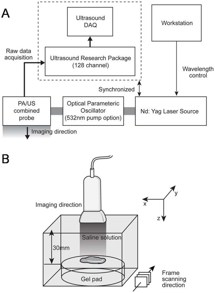
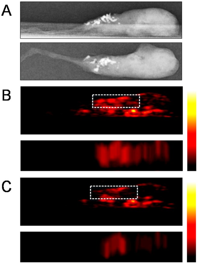
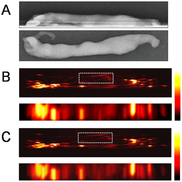
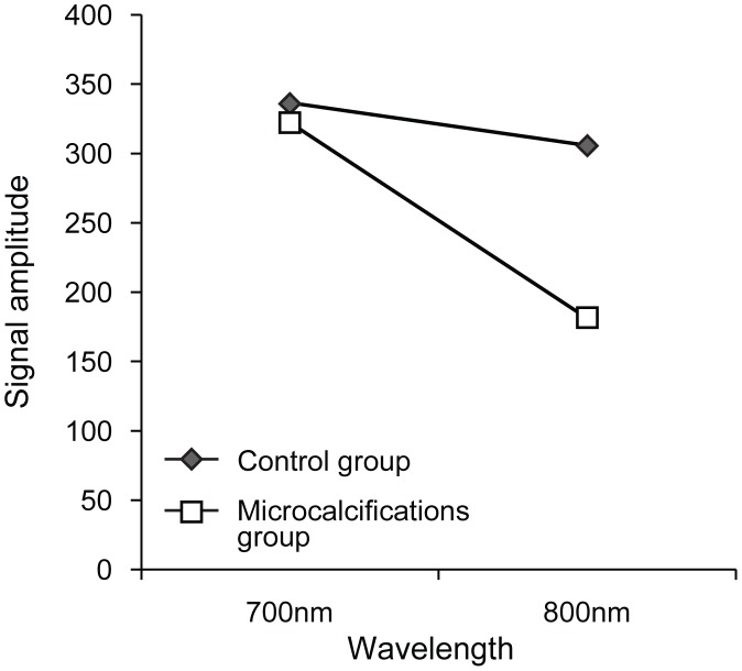
We collected 21 cores containing microcalcifications (n = 11, microcalcification group) and none (n = 10, control group) in stereotactic or ultrasound (US) guided 8-gauge vacuum-assisted biopsies. Photoacoustic (PA) images were acquired through ex vivo experiments by transmitting laser pulses with two different wavelengths (700 nm and 800 nm). The presence of microcalcifications in PA images were blindly assessed by two radiologists and compared with specimen mammography. A ratio of the signal amplitude occurring at 700 nm to that occurring at 800 nm was calculated for each PA focus and was called the PAI ratio.

RESULTS

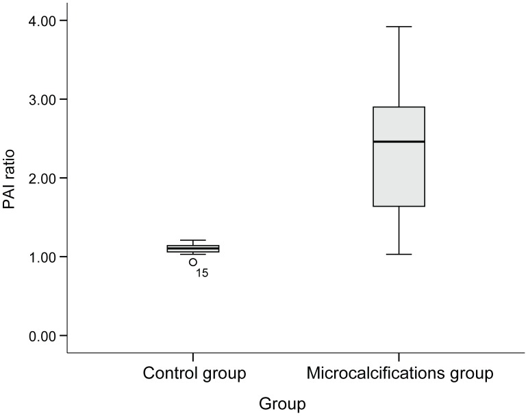
Based on the change of PA signal amplitude between 700 nm and 800 nm, 10 out of 11 specimens containing microcalcifications and 8 out of 10 specimens without calcifications were correctly identified on blind review; the sensitivity, specificity, accuracy, positive predictive and negative predictive values of our blind review were 90.91%, 80.0%, 85.71%, 83.33% and 88.89%. The PAI ratio in the microcalcification group was significantly higher than that in the control group (the median PAI ratio, 2.46 versus 1.11, respectively, P =  .001). On subgroup analysis in the microcalcification group, neither malignant diagnosis nor the number or size of calcification-foci was proven to contribute to PAI ratios.

CONCLUSION

Breast microcalcifications generated distinguishable PA signals unlike breast tissue without calcifications. So, PAI, a non-ionizing and non-invasive hybrid imaging technique, can be an alternative in overcoming the limitations of conventional US imaging.

摘要

背景

我们展示了光声成像(PAI)工具,并评估乳腺组织中的微钙化是否能在光声(PA)图像上被检测到。

方法

我们在立体定向或超声(US)引导下的8号真空辅助活检中收集了21个样本芯,其中11个含有微钙化(微钙化组),10个不含微钙化(对照组)。通过体外实验,用两种不同波长(700nm和800nm)的激光脉冲传输来获取光声(PA)图像。两名放射科医生对PA图像中微钙化的存在进行盲法评估,并与标本乳腺X线摄影进行比较。计算每个PA焦点在700nm处出现的信号幅度与在800nm处出现的信号幅度之比,称为PAI比值。

结果

基于700nm和800nm之间PA信号幅度的变化,在盲法评估中,11个含微钙化的标本中有10个、10个无钙化的标本中有8个被正确识别;我们盲法评估的敏感性、特异性、准确性、阳性预测值和阴性预测值分别为90.91%、80.0%、85.71%、83.33%和88.89%。微钙化组的PAI比值显著高于对照组(PAI比值中位数分别为2.46和1.11,P = 0.001)。在微钙化组的亚组分析中,恶性诊断以及钙化灶的数量或大小均未被证明对PAI比值有影响。

结论

乳腺微钙化产生的PA信号与无钙化的乳腺组织不同。因此,PAI作为一种非电离、非侵入性的混合成像技术,可作为克服传统超声成像局限性的一种替代方法。

https://cdn.ncbi.nlm.nih.gov/pmc/blobs/0c73/4143349/a670db20c8e9/pone.0105878.g005.jpg
https://cdn.ncbi.nlm.nih.gov/pmc/blobs/0c73/4143349/197ce0d2edbf/pone.0105878.g001.jpg
https://cdn.ncbi.nlm.nih.gov/pmc/blobs/0c73/4143349/797637721b44/pone.0105878.g002.jpg
https://cdn.ncbi.nlm.nih.gov/pmc/blobs/0c73/4143349/fd3642bdb4b5/pone.0105878.g003.jpg
https://cdn.ncbi.nlm.nih.gov/pmc/blobs/0c73/4143349/0a604acfe60c/pone.0105878.g004.jpg
https://cdn.ncbi.nlm.nih.gov/pmc/blobs/0c73/4143349/a670db20c8e9/pone.0105878.g005.jpg

相似文献

1
Photoacoustic imaging of breast microcalcifications: a preliminary study with 8-gauge core-biopsied breast specimens.乳腺微钙化的光声成像:对8号针芯活检乳腺标本的初步研究
PLoS One. 2014 Aug 25;9(8):e105878. doi: 10.1371/journal.pone.0105878. eCollection 2014.
2
Ex vivo estimation of photoacoustic imaging for detecting thyroid microcalcifications.用于检测甲状腺微钙化的光声成像的体外评估
PLoS One. 2014 Nov 21;9(11):e113358. doi: 10.1371/journal.pone.0113358. eCollection 2014.
3
Photoacoustic imaging of breast microcalcifications: a validation study with 3-dimensional ex vivo data and spectrophotometric measurement.乳腺微钙化的光声成像:一项基于三维离体数据和分光光度测量的验证研究
J Biophotonics. 2015 Jan;8(1-2):71-80. doi: 10.1002/jbio.201300100. Epub 2013 Sep 2.
4
Retrieval rate and accuracy of ultrasound-guided 14-G semi-automated core needle biopsy of breast microcalcifications.超声引导下 14-G 半自动核心针活检乳腺微钙化的检出率和准确性。
Korean J Radiol. 2014 Jan-Feb;15(1):12-9. doi: 10.3348/kjr.2014.15.1.12. Epub 2014 Jan 8.
5
Causes of failure in removing calcium in microcalcification-only lesions using 11-gauge stereotactic vacuum-assisted breast biopsy.使用 11 号活检枪立体定位真空辅助乳腺活检切除微钙化病变中钙失败的原因。
Diagn Interv Radiol. 2012 Jul-Aug;18(4):354-9. doi: 10.4261/1305-3825.DIR.5024-11.1. Epub 2012 Apr 5.
6
US-guided vacuum-assisted biopsy of microcalcifications in breast lesions and long-term follow-up results.超声引导下乳腺病变微钙化灶的真空辅助活检及长期随访结果
Korean J Radiol. 2008 Nov-Dec;9(6):503-9. doi: 10.3348/kjr.2008.9.6.503.
7
Sonographic detection and sonographically guided biopsy of breast microcalcifications.乳腺微钙化的超声检测及超声引导下活检
AJR Am J Roentgenol. 2003 Apr;180(4):941-8. doi: 10.2214/ajr.180.4.1800941.
8
Application of Raman spectroscopy to identify microcalcifications and underlying breast lesions at stereotactic core needle biopsy.拉曼光谱在立体定向核心针活检中识别微钙化及潜在乳腺病变中的应用。
Cancer Res. 2013 Jun 1;73(11):3206-15. doi: 10.1158/0008-5472.CAN-12-2313.
9
Stereotactic breast biopsy of clustered microcalcifications with a directional, vacuum-assisted device.使用定向真空辅助装置对簇状微钙化灶进行立体定向乳腺活检。
Radiology. 1997 Aug;204(2):575-6. doi: 10.1148/radiology.204.2.9240556.
10
[The demonstration of microcalcification in breast core biopsies: DIMA specimen radiography (7x) compared to the histopathological findings].[乳腺粗针活检中微钙化的显示:DIMA标本射线照相(7倍)与组织病理学结果的比较]
Rofo. 1999 Apr;170(4):347-50. doi: 10.1055/s-2007-1011052.

引用本文的文献

1
Advances in Photoacoustic Imaging of Breast Cancer.乳腺癌光声成像的进展
Sensors (Basel). 2025 Aug 5;25(15):4812. doi: 10.3390/s25154812.
2
Spatial resolution and reconstructed size accuracy using advanced beamformers in linear array-based PAT systems.基于线性阵列的光声断层成像(PAT)系统中使用先进波束形成器的空间分辨率和重建尺寸精度。
Photoacoustics. 2023 Nov 29;34:100576. doi: 10.1016/j.pacs.2023.100576. eCollection 2023 Dec.
3
Whole-Body Photoacoustic Imaging Techniques for Preclinical Small Animal Studies.用于临床前小动物研究的全身光声成像技术。

本文引用的文献

1
Intravascular optical-resolution photoacoustic tomography with a 1.1 mm diameter catheter.血管内光学分辨率光声断层成像术,采用直径 1.1 毫米的导管。
PLoS One. 2014 Mar 20;9(3):e92463. doi: 10.1371/journal.pone.0092463. eCollection 2014.
2
Imaging of an inflammatory injury in the newborn rat brain with photoacoustic tomography.新生大鼠脑部炎症性损伤的光声断层成像
PLoS One. 2013 Dec 26;8(12):e83045. doi: 10.1371/journal.pone.0083045. eCollection 2013.
3
Photoacoustic imaging of breast microcalcifications: a validation study with 3-dimensional ex vivo data and spectrophotometric measurement.
Sensors (Basel). 2022 Jul 8;22(14):5130. doi: 10.3390/s22145130.
4
The emerging role of photoacoustic imaging in clinical oncology.光声成像在临床肿瘤学中的新兴作用。
Nat Rev Clin Oncol. 2022 Jun;19(6):365-384. doi: 10.1038/s41571-022-00615-3. Epub 2022 Mar 23.
5
Transcranial photoacoustic imaging of NMDA-evoked focal circuit dynamics in the rat hippocampus.大鼠海马 NMDA 诱发的局域环路动力学的经颅光声成像。
J Neural Eng. 2020 Apr 8;17(2):025001. doi: 10.1088/1741-2552/ab78ca.
6
State-of-the-Art Preclinical Photoacoustic Imaging in Oncology: Recent Advances in Cancer Theranostics.肿瘤学中先进的临床前光声成像:癌症治疗诊断学的最新进展。
Contrast Media Mol Imaging. 2019 Apr 30;2019:5080267. doi: 10.1155/2019/5080267. eCollection 2019.
7
Intraoperative photoacoustic screening of breast cancer: a new perspective on malignancy visualization and surgical guidance.术中光声筛查乳腺癌:一种用于可视化恶性肿瘤和指导手术的新方法。
J Biomed Opt. 2019 May;24(5):1-12. doi: 10.1117/1.JBO.24.5.056002.
8
Multimodal photoacoustic imaging as a tool for sentinel lymph node identification and biopsy guidance.多模态光声成像作为前哨淋巴结识别和活检引导的工具。
Biomed Eng Lett. 2018 Apr 21;8(2):183-191. doi: 10.1007/s13534-018-0068-1. eCollection 2018 May.
9
Photoacoustic Imaging for Differential Diagnosis of Benign Polyps versus Malignant Polyps of the Gallbladder: A Preliminary Study.用于胆囊良性息肉与恶性息肉鉴别诊断的光声成像:一项初步研究。
Korean J Radiol. 2017 Sep-Oct;18(5):821-827. doi: 10.3348/kjr.2017.18.5.821. Epub 2017 Jul 17.
10
Programmable Real-time Clinical Photoacoustic and Ultrasound Imaging System.可编程实时临床光声与超声成像系统
Sci Rep. 2016 Oct 12;6:35137. doi: 10.1038/srep35137.
乳腺微钙化的光声成像:一项基于三维离体数据和分光光度测量的验证研究
J Biophotonics. 2015 Jan;8(1-2):71-80. doi: 10.1002/jbio.201300100. Epub 2013 Sep 2.
4
New image processing technique for evaluating breast microcalcifications: a comparative study.用于评估乳腺微钙化的新型图像处理技术:一项对比研究。
J Ultrasound Med. 2012 Jun;31(6):885-93. doi: 10.7863/jum.2012.31.6.885.
5
Enhancement of photoacoustic image quality by sound speed correction: ex vivo evaluation.通过声速校正提高光声图像质量:离体评估
Opt Express. 2012 Jan 30;20(3):3082-90. doi: 10.1364/OE.20.003082.
6
Raman spectroscopy: a real-time tool for identifying microcalcifications during stereotactic breast core needle biopsies.拉曼光谱法:一种在立体定向乳腺粗针活检过程中识别微钙化的实时工具。
Biomed Opt Express. 2011 Oct 1;2(10):2792-803. doi: 10.1364/BOE.2.002792. Epub 2011 Sep 14.
7
Photoacoustic angiography of the breast.乳腺光声血管造影。
Med Phys. 2010 Nov;37(11):6096-100. doi: 10.1118/1.3497677.
8
Sonographic findings of high-grade and non-high-grade ductal carcinoma in situ of the breast.乳腺高级别和非高级别导管原位癌的超声表现。
J Ultrasound Med. 2010 Dec;29(12):1687-97. doi: 10.7863/jum.2010.29.12.1687.
9
New relationships between breast microcalcifications and cancer.乳腺微钙化与癌症的新关系。
Br J Cancer. 2010 Sep 28;103(7):1034-9. doi: 10.1038/sj.bjc.6605873. Epub 2010 Sep 14.
10
Radiation doses and cancer risks from breast imaging studies.乳腺影像学研究的辐射剂量与癌症风险。
Radiology. 2010 Oct;257(1):246-53. doi: 10.1148/radiol.10100570. Epub 2010 Aug 24.