Barow Ewgenia, Neumann Wolf-Julian, Brücke Christof, Huebl Julius, Horn Andreas, Brown Peter, Krauss Joachim K, Schneider Gerd-Helge, Kühn Andrea A
Department of Neurology, Campus Virchow Klinikum, Charité-University Medicine Berlin, Berlin, Germany.
Nuffield Department of Clinical Neurosciences, University of Oxford, Oxford, UK.
Brain. 2014 Nov;137(Pt 11):3012-3024. doi: 10.1093/brain/awu258. Epub 2014 Sep 10.
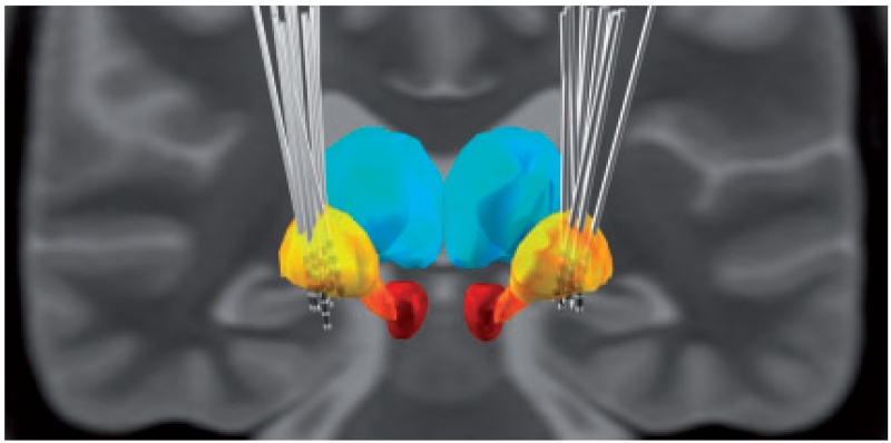
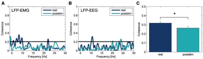
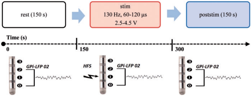
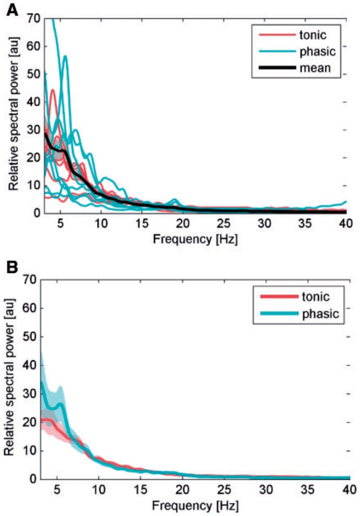
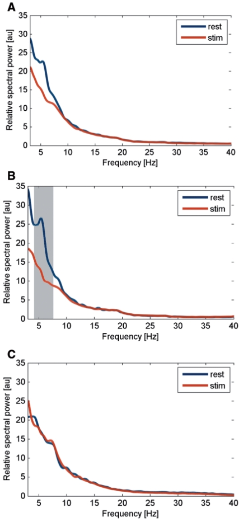
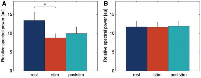
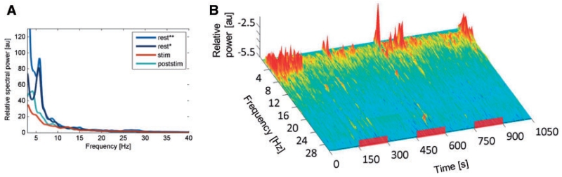
Deep brain stimulation of the globus pallidus internus alleviates involuntary movements in patients with dystonia. However, the mechanism is still not entirely understood. One hypothesis is that deep brain stimulation suppresses abnormally enhanced synchronized oscillatory activity within the motor cortico-basal ganglia network. Here, we explore deep brain stimulation-induced modulation of pathological low frequency (4-12 Hz) pallidal activity that has been described in local field potential recordings in patients with dystonia. Therefore, local field potentials were recorded from 16 hemispheres in 12 patients undergoing deep brain stimulation for severe dystonia using a specially designed amplifier allowing simultaneous high frequency stimulation at therapeutic parameter settings and local field potential recordings. For coherence analysis electroencephalographic activity (EEG) over motor areas and electromyographic activity (EMG) from affected neck muscles were recorded before and immediately after cessation of high frequency stimulation. High frequency stimulation led to a significant reduction of mean power in the 4-12 Hz band by 24.8 ± 7.0% in patients with predominantly phasic dystonia. A significant decrease of coherence between cortical EEG and pallidal local field potential activity in the 4-12 Hz range was revealed for the time period of 30 s after switching off high frequency stimulation. Coherence between EMG activity and pallidal activity was mainly found in patients with phasic dystonic movements where it was suppressed after high frequency stimulation. Our findings suggest that high frequency stimulation may suppress pathologically enhanced low frequency activity in patients with phasic dystonia. These dystonic features are the quickest to respond to high frequency stimulation and may thus directly relate to modulation of pathological basal ganglia activity, whereas improvement in tonic features may depend on long-term plastic changes within the motor network.
苍白球内侧部的深部脑刺激可缓解肌张力障碍患者的不自主运动。然而,其机制仍未完全明确。一种假说是,深部脑刺激可抑制运动皮质-基底神经节网络内异常增强的同步振荡活动。在此,我们探讨深部脑刺激对肌张力障碍患者局部场电位记录中所描述的病理性低频(4 - 12赫兹)苍白球活动的调节作用。因此,使用专门设计的放大器,在12例因严重肌张力障碍接受深部脑刺激的患者的16个半球记录局部场电位,该放大器允许在治疗参数设置下同时进行高频刺激和局部场电位记录。为进行相干分析,在高频刺激停止前和停止后立即记录运动区域的脑电图活动(EEG)以及患侧颈部肌肉的肌电图活动(EMG)。高频刺激导致以相位性肌张力障碍为主的患者在4 - 12赫兹频段的平均功率显著降低24.8±7.0%。在高频刺激关闭后的30秒内,皮质EEG与苍白球局部场电位活动在4 - 12赫兹范围内的相干性显著降低。EMG活动与苍白球活动之间的相干性主要见于有相位性肌张力障碍运动的患者,高频刺激后其相干性受到抑制。我们的研究结果表明,高频刺激可能抑制相位性肌张力障碍患者病理性增强的低频活动。这些肌张力障碍特征对高频刺激反应最快,因此可能直接与病理性基底神经节活动的调节有关,而强直性特征的改善可能取决于运动网络内的长期可塑性变化。