Park Sung Pyo, Lee Winston, Bae Eun Jin, Greenstein Vivianne, Sin Bum Ho, Chang Stanley, Tsang Stephen H
Bernard and Shirlee Brown Glaucoma Laboratory, Edward S. Harkness Eye Institute, Columbia University, 160 Fort Washington Avenue, Room 513, New York, NY 10032, USA.
Ophthalmic Surg Lasers Imaging Retina. 2014 Sep-Oct;45(5):469-473. doi: 10.3928/23258160-20140908-01.
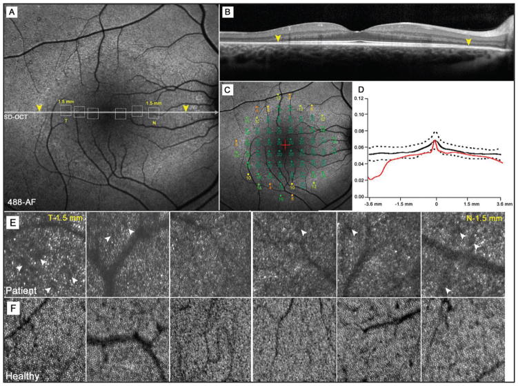
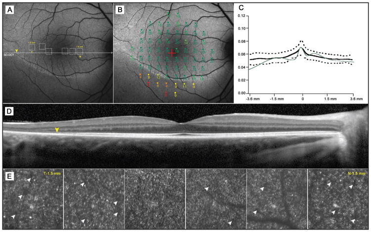
The authors report the use of adaptive-optics scanning laser ophthalmoscopy (AO-SLO) to investigate RHO, D190N autosomal-dominant retinitis pigmentosa in two siblings (11 and 16 years old, respectively). Each patient exhibited distinct hyperautofluorescence patterns in which the outer borders corresponded to inner segment ellipsoid band disruption. Areas within the hyperautofluorescence patterns exhibited normal photoreceptor outer segments and retinal pigment epithelium. However, AO-SLO imaging revealed noticeable spacing irregularities in the cone mosaic. AO-SLO allows researchers to characterize retinal structural abnormalities with precision so that early structural changes in retinitis pigmentosa can be identified and reconciled with genetic findings.
作者报告了使用自适应光学扫描激光检眼镜(AO-SLO)对两名分别为11岁和16岁的兄弟姐妹进行RHO D190N常染色体显性遗传性视网膜色素变性的研究。每位患者均表现出独特的高自发荧光模式,其外边界对应于内节椭圆体带中断。高自发荧光模式内的区域显示光感受器外节和视网膜色素上皮正常。然而,AO-SLO成像显示视锥细胞镶嵌存在明显的间距不规则。AO-SLO使研究人员能够精确地描述视网膜结构异常,从而可以识别视网膜色素变性的早期结构变化并与基因研究结果相匹配。