Muricy Edna Cleide Mendes, Lemes Romilda Aparecida, Bombarda Sidney, Ferrazoli Lucilaine, Chimara Erica
Medical Mycology Laboratory (LIM-53), Instituto de Medicina Tropical, São Paulo, SP, Brazil.
Tuberculosis and Mycobacteriosis Branch, Instituto Adolfo Lutz, São Paulo, SP, Brazil.
Rev Inst Med Trop Sao Paulo. 2014 Sep-Oct;56(5):397-401. doi: 10.1590/s0036-46652014000500005.
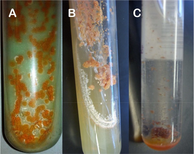
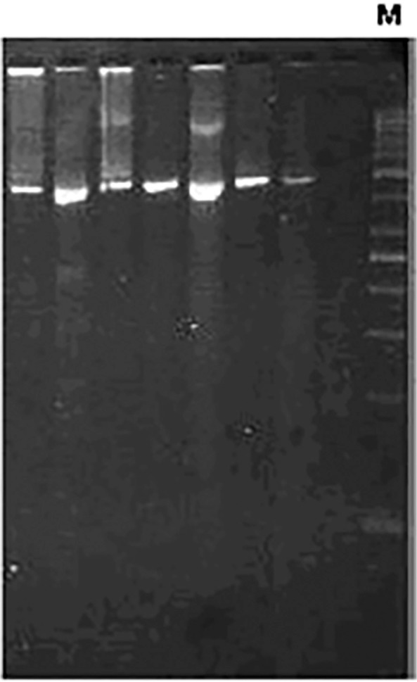
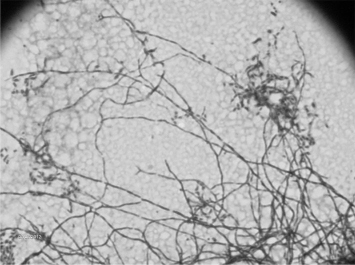
New methodologies were developed for the identification of Nocardia but the initial diagnosis still requires a fast and accurate method, mainly due to the similarity to Mycobacterium, both clinical and bacteriologically. Growth on Löwenstein-Jensen (LJ) medium, presence of acid-fast bacilli through Ziehl-Neelsen staining, and colony morphology can be confusing aspects between Nocardia and Mycobacterium. This study describes the occurrence of Nocardia spp. in a mycobacterial-reference laboratory, observing the main difficulties in differentiating Nocardia spp. from Mycobacterium spp., and correlating isolates with nocardiosis cases. Laboratory records for the period between 2008 and 2012 were analyzed, and the isolates identified as Nocardia sp. or as non-acid-fast filamentous bacilli were selected. Epidemiological and bacteriological data were analyzed as well. Thirty-three isolates identified as Nocardia sp. and 22 as non-acid-fast bacilli were selected for this study, and represented 0.12% of isolates during the study period. The presumptive identification was based on macroscopic and microscopic morphology, resistance to lysozyme and restriction profiles using the PRA-hsp65 method. Nocardia spp. can grow on media for mycobacteria isolation (LJ and BBL MGIT™) and microscopy and colony morphology are very similar to some mycobacteria species. Seventeen patients (54.8%) were reported and treated for tuberculosis, but presented signs and symptoms of nocardiosis. It was concluded that the occurrence of Nocardia sp. during the study period was 0.12%. Isolates with characteristics of filamentous bacilli, forming aerial hyphae, with colonies that may be pigmented, rough and without the BstEII digestion pattern in PRA-hsp65 method are suggestive of Nocardia spp. For a mycobacterial routine laboratory, a flow for the presumptive identification of Nocardia is essential, allowing the use of more accurate techniques for the correct identification, proper treatment and better quality of life for patients.
已开发出用于鉴定诺卡菌的新方法,但初始诊断仍需要一种快速准确的方法,这主要是因为在临床和细菌学方面,诺卡菌与分枝杆菌相似。在罗氏培养基(LJ)上生长、通过萋尼染色显示抗酸杆菌以及菌落形态,这些在诺卡菌和分枝杆菌之间可能会造成混淆。本研究描述了诺卡菌属在一个分枝杆菌参考实验室中的出现情况,观察了区分诺卡菌属与分枝杆菌属的主要困难,并将分离株与诺卡菌病病例相关联。分析了2008年至2012年期间的实验室记录,并选择了鉴定为诺卡菌属或非抗酸丝状杆菌的分离株。还分析了流行病学和细菌学数据。本研究选择了33株鉴定为诺卡菌属的分离株和22株非抗酸杆菌,占研究期间分离株的0.12%。初步鉴定基于宏观和微观形态、对溶菌酶的抗性以及使用PRA-hsp65方法的限制性图谱。诺卡菌属可在用于分枝杆菌分离的培养基(LJ和BBL MGIT™)上生长,其显微镜检查和菌落形态与某些分枝杆菌种类非常相似。有17名患者(54.8%)被报告患有结核病并接受治疗,但表现出诺卡菌病的体征和症状。得出的结论是,在研究期间诺卡菌属的出现率为0.12%。具有丝状杆菌特征、形成气生菌丝、菌落可能有色素沉着、粗糙且在PRA-hsp65方法中无BstEII消化模式的分离株提示为诺卡菌属。对于分枝杆菌常规实验室而言,诺卡菌的初步鉴定流程至关重要,这能让更准确的技术得以用于正确鉴定、恰当治疗并提高患者的生活质量。