Kim Eun Jin, Choi Joo Yeon, Park Byung Cheol, Lee Bog-Hieu
Department of Food and Nutrition, Graduate School of Chung-Ang University, Seoul 156-756, Korea.
Department of Dermatology, College of Medicine, Dankook University, Chungnam 330-714, Korea.
Prev Nutr Food Sci. 2014 Sep;19(3):136-44. doi: 10.3746/pnf.2014.19.3.136.
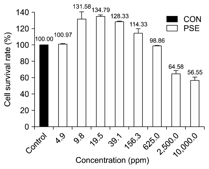
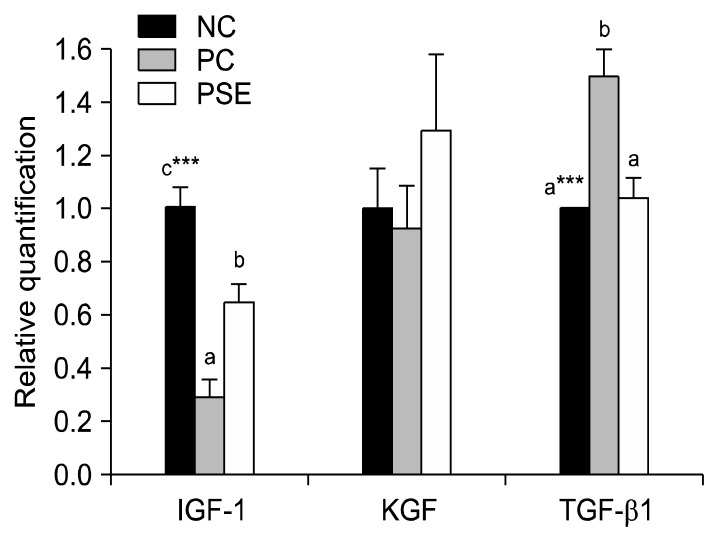
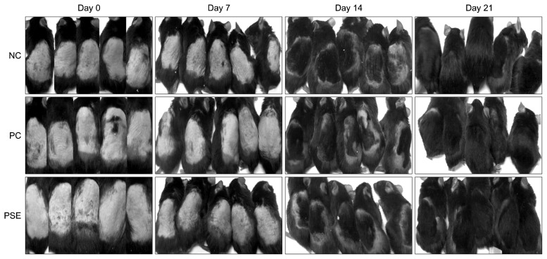
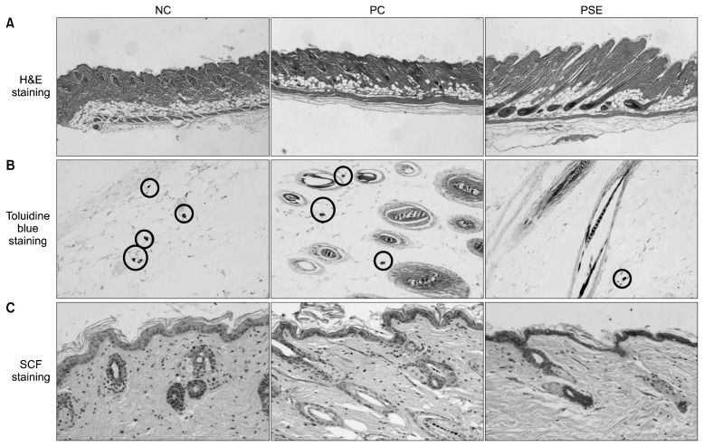
This study was conducted to evaluate the effects of Platycarya strobilacea S. et Z. (PSE) extract on mouse hair growth and to determine the mechanism of action of PSE. PSE was purchased and its antioxidant activities, such as electron donating ability, total polyphenol content, and flavonoid content were tested. Toxicity during topical treatment was determined by the CCK-8 assay, a cell viability test. Fifteen 4-week-old male C57BL/6 mice were assigned to receive one of three treatments: dimethyl sulfoxide (negative control), minoxidil (positive control) or PSE. Test materials were topically applied to the shaved dorsal skin of each mouse daily for 3 weeks. After 21 days, we observed skin tissue hair follicle morphology and length, mast cell number, and stem cell factor (SCF) expression using hematoxylin and eosin (H&E), toluidine blue, and immunohistochemical staining, respectively. Furthermore, the expression of cytokines involved in hair growth [i.e., insulin-like growth factor (IGF)-1, keratinocyte growth factor (KGF), and transforming growth factor (TGF)-β1] was determined by PCR. PSE was found to have very high antioxidant activity. The cell viability rate of PSE-treated mice was markedly higher than that of mice in the control group. We also observed an increase in hair follicle length, strong SCF staining, and a decrease in mast cell number in the PSE group. In addition, PSE-treated mice had higher IGF-1 and KGF expression and lower TGF-β1 expression than mice in the minoxidil-treated group. These results suggest that topical application of PSE promotes hair growth by intensifying SCF, suppressing mast cell production, and increasing hair growth-promoting cytokine expression.
本研究旨在评估化香树提取物(PSE)对小鼠毛发生长的影响,并确定其作用机制。购买了PSE并测试了其抗氧化活性,如电子供体能力、总多酚含量和类黄酮含量。通过CCK-8试验(一种细胞活力测试)确定局部治疗期间的毒性。将15只4周龄雄性C57BL/6小鼠分为三组接受不同处理:二甲亚砜(阴性对照)、米诺地尔(阳性对照)或PSE。将测试材料每天局部涂抹于每只小鼠剃毛的背部皮肤,持续3周。21天后,我们分别使用苏木精和伊红(H&E)染色、甲苯胺蓝染色和免疫组织化学染色观察皮肤组织毛囊形态和长度、肥大细胞数量以及干细胞因子(SCF)表达。此外,通过PCR测定参与毛发生长的细胞因子[即胰岛素样生长因子(IGF)-1、角质形成细胞生长因子(KGF)和转化生长因子(TGF)-β1]的表达。发现PSE具有非常高的抗氧化活性。PSE处理组小鼠的细胞活力率明显高于对照组小鼠。我们还观察到PSE组毛囊长度增加、SCF染色增强以及肥大细胞数量减少。此外,与米诺地尔处理组小鼠相比,PSE处理组小鼠的IGF-1和KGF表达更高,而TGF-β1表达更低。这些结果表明,局部应用PSE通过增强SCF、抑制肥大细胞产生以及增加促进毛发生长的细胞因子表达来促进毛发生长。