Camerlingo Carlo, d'Apuzzo Fabrizia, Grassia Vincenzo, Perillo Letizia, Lepore Maria
CNR-SPIN, Istituto Superconduttori, Materiali Innovativi e Dispositivi, via Campi Flegrei 34, Pozzuoli 80078, Italy.
Dip. Multidisciplinare di Specialità Medico-Chirurgiche e Odontoiatriche, Seconda Università di Napoli, via L. De Crecchio 6, Napoli 80138, Italy.
Sensors (Basel). 2014 Nov 27;14(12):22552-63. doi: 10.3390/s141222552.
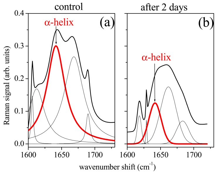
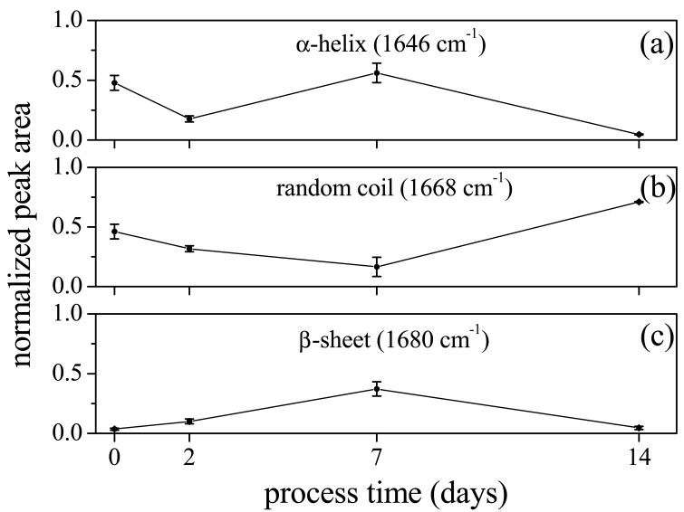
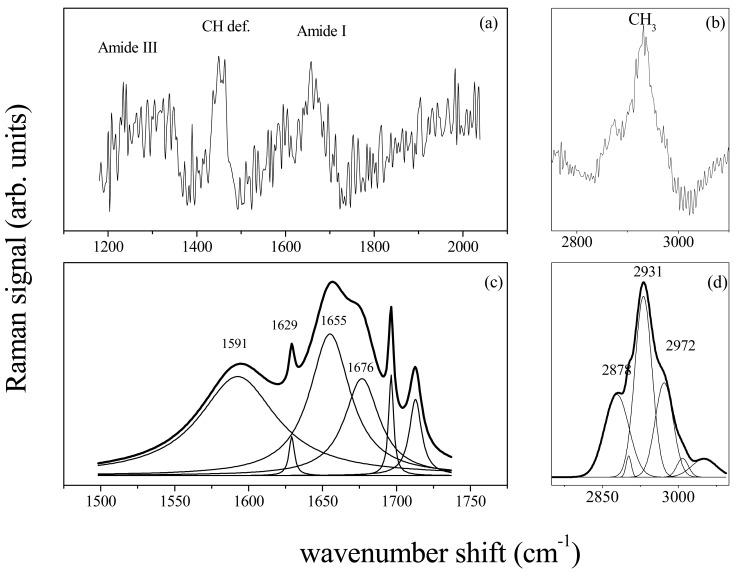
Micro-Raman Spectroscopy is an efficient method for analyzing biological specimens due to its sensitivity to subtle chemical and structural changes. The aim of this study was to use micro-Raman spectroscopy to analyze chemical and structural changes in periodontal ligament after orthodontic force application and in gingival crevicular fluid in presence of periodontal disease. The biopsy of periodontal ligament samples of premolars extracted for orthodontic reasons and the gingival crevicular fluid samples collected by using absorbent paper cones; were analyzed by micro-Raman spectroscopy. Changes of the secondary protein structure related to different times of orthodontic force application were reported; whereas an increase of carotene was revealed in patients affected by periodontal inflammation.
显微拉曼光谱法因其对细微化学和结构变化的敏感性,是分析生物样本的一种有效方法。本研究的目的是使用显微拉曼光谱法分析正畸力施加后牙周膜以及存在牙周疾病时龈沟液中的化学和结构变化。对因正畸原因拔除的前磨牙的牙周膜样本进行活检,并使用吸水纸锥收集龈沟液样本;通过显微拉曼光谱法进行分析。报告了与正畸力施加不同时间相关的二级蛋白质结构变化;而在患有牙周炎症的患者中发现胡萝卜素增加。