Pflanzer R, Hofmann M, Shelke A, Habib A, Derwich W, Schmitz-Rixen T, Bernd A, Kaufmann R, Bereiter-Hahn J
Dept. of Dermatology, Venerology and Allergology, Goethe University Frankfurt, 60590 Frankfurt/Main, Germany.
Dept. of Dermatology, Venerology and Allergology, Goethe University Frankfurt, 60590 Frankfurt/Main, Germany.
Transl Oncol. 2014 Dec;7(6):681-6. doi: 10.1016/j.tranon.2014.09.013.
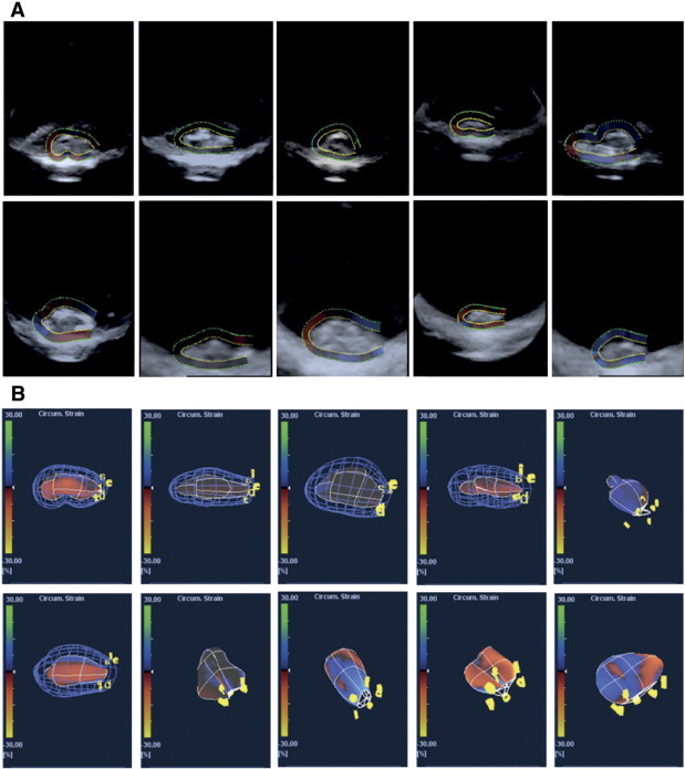
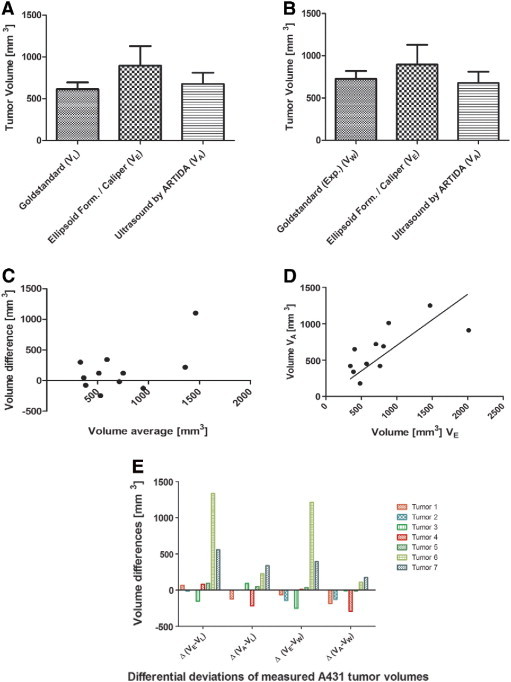
Determination of tumor volume in subcutaneously inoculated xenograft models is a standard procedure for clinical and preclinical evaluation of tumor response to treatment. Practitioners frequently use a hands-on caliper method in conjunction with a simplified formula to assess tumor volume. Non-invasive and more precise techniques as investigation by MR or (μ)CT exist but come with various adverse effects in terms of radiation, complex setup or elevated cost of investigations. Therefore, we propose an advanced three-dimensional sonographic imaging technique to determine small tumor volumes in xenografts with high precision and minimized observer variability. We present a study on xenograft carcinoma tumors from which volumes and shapes were calculated with the standard caliper method as well as with a clinically available three-dimensional ultrasound scanner and subsequent processing software. Statistical analysis reveals the suitability of this non-invasive approach for the purpose of a quick and precise calculation of tumor volume in small rodents.
在皮下接种的异种移植模型中测定肿瘤体积是临床和临床前评估肿瘤对治疗反应的标准程序。从业者经常使用手动卡尺法并结合简化公式来评估肿瘤体积。虽然存在如磁共振成像(MR)或微计算机断层扫描(μCT)等非侵入性且更精确的技术,但这些技术在辐射、设置复杂或检查成本高等方面存在各种不利影响。因此,我们提出一种先进的三维超声成像技术,以高精度且最小化观察者变异性来测定异种移植中小肿瘤的体积。我们展示了一项关于异种移植癌肿瘤的研究,通过标准卡尺法以及临床可用的三维超声扫描仪和后续处理软件计算肿瘤的体积和形状。统计分析表明这种非侵入性方法适用于快速、精确地计算小型啮齿动物的肿瘤体积。