Chang Kelly C, Bayer Jason D, Trayanova Natalia A
Institute for Computational Medicine, Department of Biomedical Engineering, Johns Hopkins University, Baltimore, Maryland, United States of America.
IHU-LIRYC - L'Institut de RYthmologie et Modélisation Cardiaque, University of Bordeaux, Bordeaux, France.
PLoS Comput Biol. 2014 Dec 11;10(12):e1004011. doi: 10.1371/journal.pcbi.1004011. eCollection 2014 Dec.
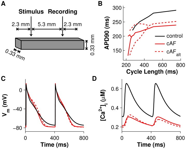
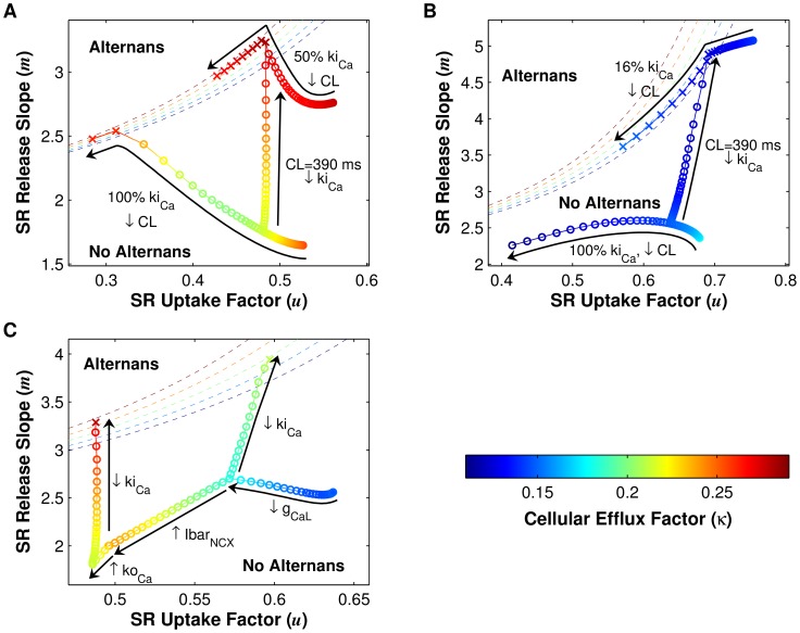
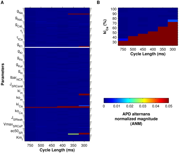
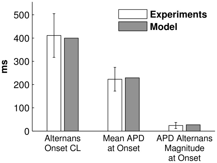
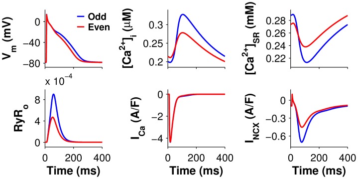
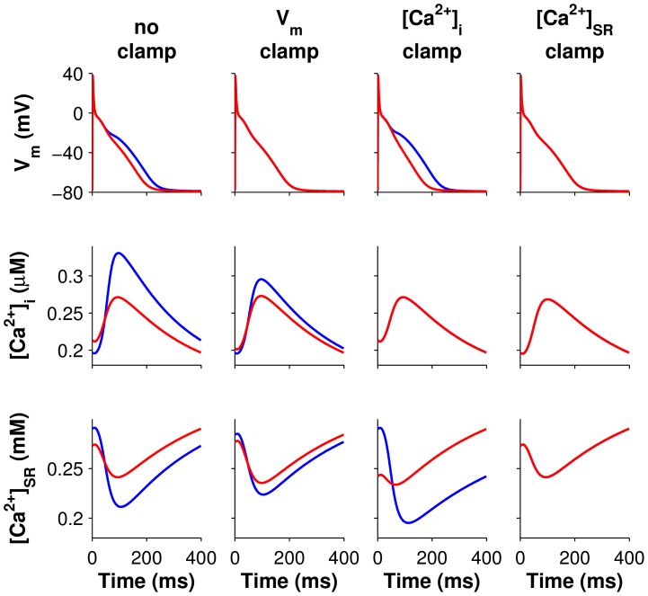
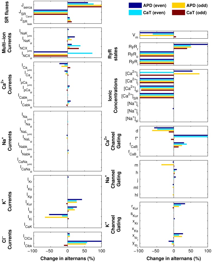
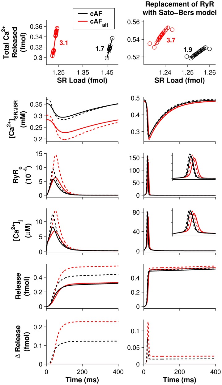
Atrial fibrillation (AF) is the most common cardiac arrhythmia, but our knowledge of the arrhythmogenic substrate is incomplete. Alternans, the beat-to-beat alternation in the shape of cardiac electrical signals, typically occurs at fast heart rates and leads to arrhythmia. However, atrial alternans have been observed at slower pacing rates in AF patients than in controls, suggesting that increased vulnerability to arrhythmia in AF patients may be due to the proarrythmic influence of alternans at these slower rates. As such, alternans may present a useful therapeutic target for the treatment and prevention of AF, but the mechanism underlying alternans occurrence in AF patients at heart rates near rest is unknown. The goal of this study was to determine how cellular changes that occur in human AF affect the appearance of alternans at heart rates near rest. To achieve this, we developed a computational model of human atrial tissue incorporating electrophysiological remodeling associated with chronic AF (cAF) and performed parameter sensitivity analysis of ionic model parameters to determine which cellular changes led to alternans. Of the 20 parameters tested, only decreasing the ryanodine receptor (RyR) inactivation rate constant (kiCa) produced action potential duration (APD) alternans seen clinically at slower pacing rates. Using single-cell clamps of voltage, fluxes, and state variables, we determined that alternans onset was Ca2+-driven rather than voltage-driven and occurred as a result of decreased RyR inactivation which led to increased steepness of the sarcoplasmic reticulum (SR) Ca2+ release slope. Iterated map analysis revealed that because SR Ca2+ uptake efficiency was much higher in control atrial cells than in cAF cells, drastic reductions in kiCa were required to produce alternans at comparable pacing rates in control atrial cells. These findings suggest that RyR kinetics may play a critical role in altered Ca2+ homeostasis which drives proarrhythmic APD alternans in patients with AF.
心房颤动(AF)是最常见的心律失常,但我们对其致心律失常基质的了解并不完整。交替变化,即心脏电信号形态上的逐搏交替,通常发生在心率较快时并导致心律失常。然而,与对照组相比,在AF患者中观察到在较慢起搏频率下就出现了心房交替变化,这表明AF患者心律失常易感性增加可能是由于这些较慢频率下交替变化的促心律失常作用。因此,交替变化可能是治疗和预防AF的一个有用的治疗靶点,但AF患者在静息心率附近出现交替变化的潜在机制尚不清楚。本研究的目的是确定人类AF中发生的细胞变化如何影响静息心率附近交替变化的出现。为实现这一目标,我们建立了一个包含与慢性AF(cAF)相关的电生理重塑的人类心房组织计算模型,并对离子模型参数进行参数敏感性分析,以确定哪些细胞变化导致了交替变化。在测试的20个参数中,只有降低兰尼碱受体(RyR)失活速率常数(kiCa)才会产生临床上在较慢起搏频率下出现的动作电位时程(APD)交替变化。通过对电压、通量和状态变量的单细胞钳制,我们确定交替变化的起始是由Ca2+驱动而非电压驱动的,并且是由于RyR失活减少导致肌浆网(SR)Ca2+释放斜率变陡而发生的。迭代映射分析表明,由于对照心房细胞中SR Ca2+摄取效率远高于cAF细胞,因此在对照心房细胞中以可比的起搏频率产生交替变化需要大幅降低kiCa。这些发现表明,RyR动力学可能在改变Ca2+稳态中起关键作用,而Ca2+稳态改变会驱动AF患者出现促心律失常的APD交替变化。