Sathitruangsak Chirawadee, Righolt Christiaan H, Klewes Ludger, Tammur Pille, Ilus Tiiu, Tamm Anu, Punab Mari, Olujohungbe Adebayo, Mai Sabine
Manitoba Institute of Cell Biology, University of Manitoba, CancerCare Manitoba, Winnipeg, Manitoba, Canada; Division of Medical Oncology, Department of Internal Medicine, Prince of Songkla University, Songkhla, Thailand.
J Cell Biochem. 2015 May;116(5):704-10. doi: 10.1002/jcb.25030.
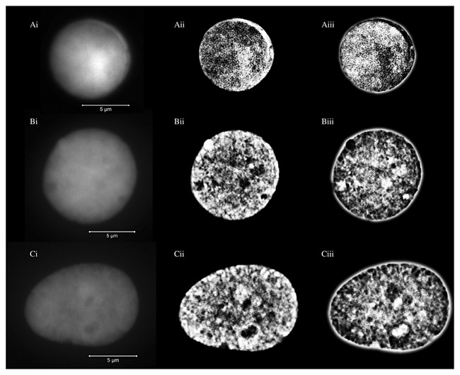
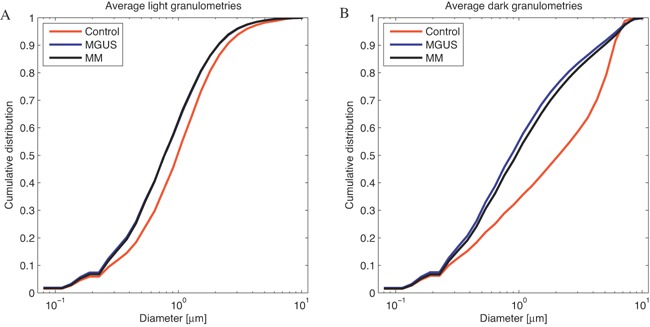
The mammalian nucleus has a distinct substructure that cannot be visualized directly by conventional microscopy. In this study, the organization of the DNA within the nucleus of multiple myeloma (MM) cells, their precursor cells (monoclonal gammopathy of undetermined significance; MGUS) and control lymphocytes of the representative patients is visualized and quantified by superresolution microscopy. Three-dimensional structured illumination microscopy (3D-SIM) increases the spatial resolution beyond the limits of conventional widefield fluorescence microscopy. 3D-SIM reveals new insights into the nuclear architecture of cancer as we show for the first time that it resolves organizational differences in intranuclear DNA organization of myeloma cells in MGUS and in MM patients. In addition, we report a significant increase in nuclear submicron DNA structure and structure of the DNA-free space in myeloma nuclei compared to normal lymphocyte nuclei. Our study provides previously unknown details of the nanoscopic DNA architecture of interphase nuclei of the normal lymphocytes, MGUS and MM cells. This study opens new avenues to understanding the disease progression from MGUS to MM.
哺乳动物的细胞核具有独特的亚结构,无法通过传统显微镜直接观察到。在本研究中,通过超分辨率显微镜对多发性骨髓瘤(MM)细胞、其前体细胞(意义未明的单克隆丙种球蛋白病;MGUS)以及代表性患者的对照淋巴细胞的细胞核内DNA组织进行了可视化和定量分析。三维结构照明显微镜(3D-SIM)将空间分辨率提高到超出传统宽场荧光显微镜的极限。3D-SIM揭示了癌症核结构的新见解,因为我们首次表明它能够分辨MGUS和MM患者骨髓瘤细胞核内DNA组织的组织差异。此外,我们报告与正常淋巴细胞核相比,骨髓瘤细胞核内亚微米级DNA结构和无DNA空间的结构显著增加。我们的研究提供了正常淋巴细胞、MGUS和MM细胞间期核纳米级DNA结构以前未知的细节。这项研究为理解从MGUS到MM的疾病进展开辟了新途径。