Delivopoulos Evangelos, Ouberai Myriam M, Coffey Paul D, Swann Marcus J, Shakesheff Kevin M, Welland Mark E
Nanoscience Centre, Department of Engineering, University of Cambridge, Cambridge CB3 0FF, UK; School of Systems Engineering, University of Reading, Reading RG6 6AY, UK.
Nanoscience Centre, Department of Engineering, University of Cambridge, Cambridge CB3 0FF, UK.
Colloids Surf B Biointerfaces. 2015 Feb 1;126:169-77. doi: 10.1016/j.colsurfb.2014.12.020. Epub 2014 Dec 16.
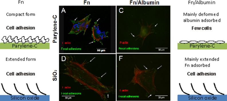
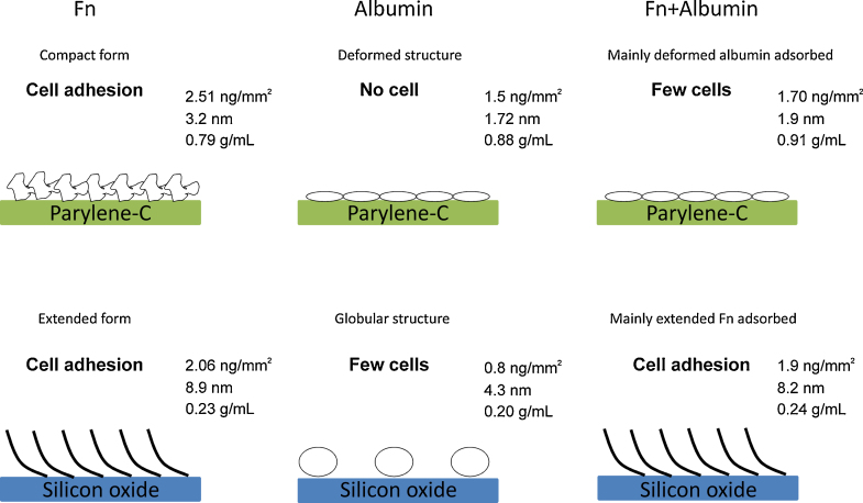
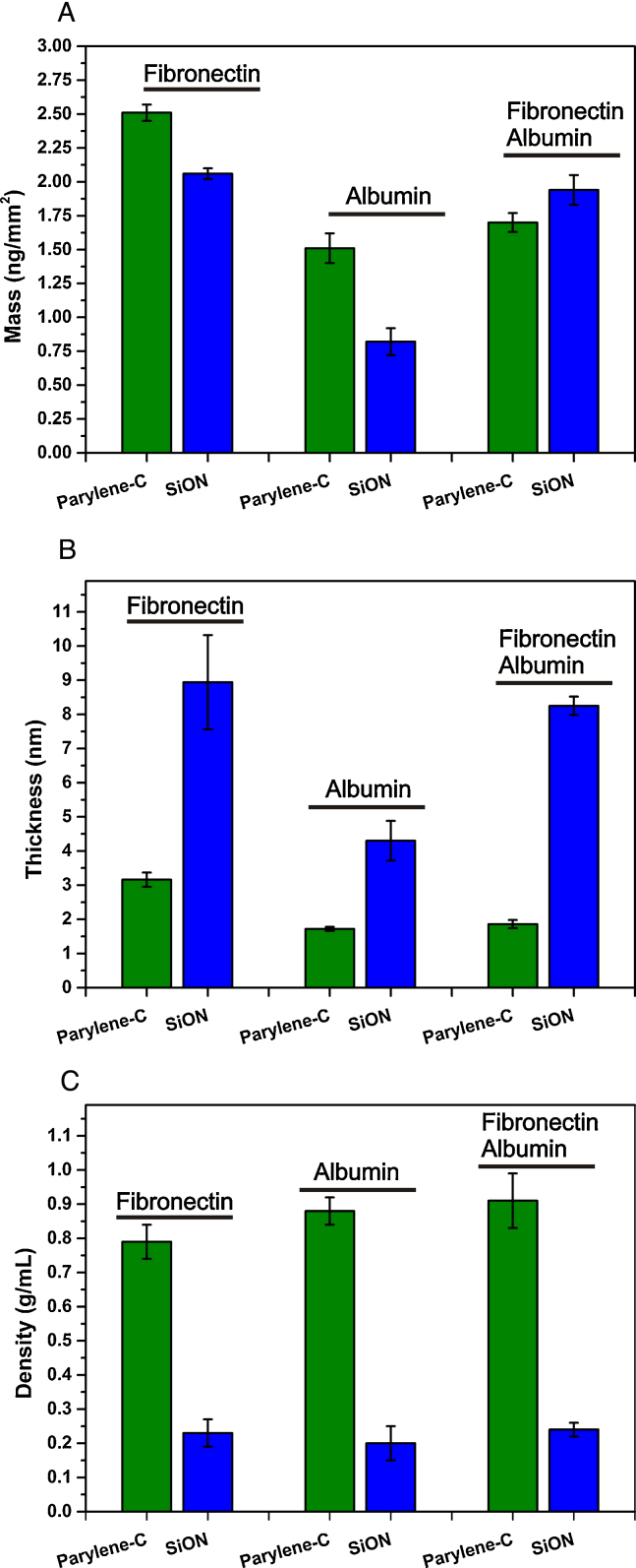
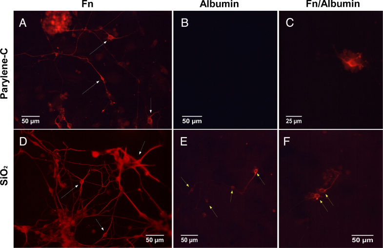
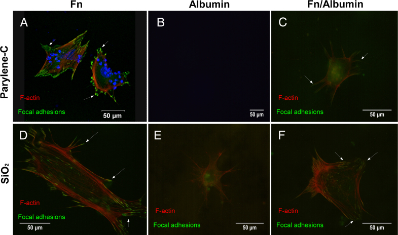
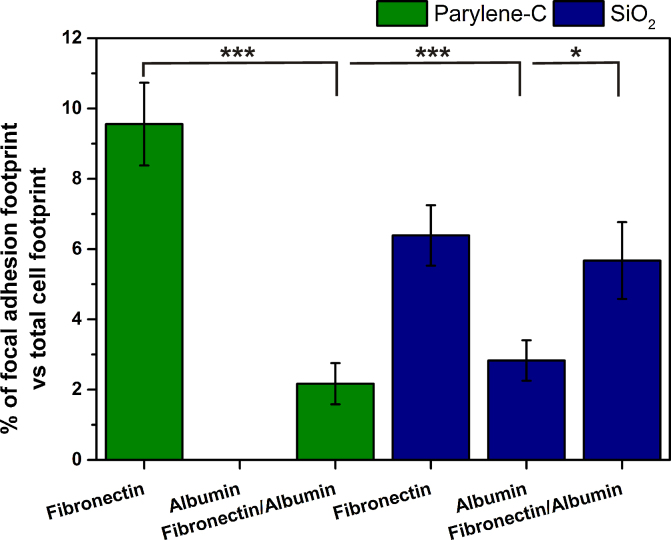
Among the range of materials used in bioengineering, parylene-C has been used in combination with silicon oxide and in presence of the serum proteins, in cell patterning. However, the structural properties of adsorbed serum proteins on these substrates still remain elusive. In this study, we use an optical biosensing technique to decipher the properties of fibronectin (Fn) and serum albumin adsorbed on parylene-C and silicon oxide substrates. Our results show the formation of layers with distinct structural and adhesive properties. Thin, dense layers are formed on parylene-C, whereas thicker, more diffuse layers are formed on silicon oxide. These results suggest that Fn acquires a compact structure on parylene-C and a more extended structure on silicon oxide. Nonetheless, parylene-C and silicon oxide substrates coated with Fn host cell populations that exhibit focal adhesion complexes and good cell attachment. Albumin adopts a deformed structure on parylene-C and a globular structure on silicon oxide, and does not support significant cell attachment on either surface. Interestingly, the co-incubation of Fn and albumin at the ratio found in serum, results in the preferential adsorption of albumin on parylene-C and Fn on silicon oxide. This finding is supported by the exclusive formation of focal adhesion complexes in differentiated mouse embryonic stem cells (CGR8), cultured on Fn/albumin coated silicon oxide, but not on parylene-C. The detailed information provided in this study on the distinct properties of layers of serum proteins on substrates such as parylene-C and silicon oxide is highly significant in developing methods for cell patterning.
在生物工程中使用的一系列材料中,聚对二甲苯-C已与氧化硅结合使用,并在血清蛋白存在的情况下用于细胞图案化。然而,这些底物上吸附的血清蛋白的结构特性仍然难以捉摸。在本研究中,我们使用光学生物传感技术来解读吸附在聚对二甲苯-C和氧化硅底物上的纤连蛋白(Fn)和血清白蛋白的特性。我们的结果表明形成了具有不同结构和粘附特性的层。在聚对二甲苯-C上形成薄而致密的层,而在氧化硅上形成更厚、更分散的层。这些结果表明,Fn在聚对二甲苯-C上获得紧密结构,在氧化硅上获得更伸展的结构。尽管如此,涂有Fn的聚对二甲苯-C和氧化硅底物支持表现出粘着斑复合物和良好细胞附着的细胞群体。白蛋白在聚对二甲苯-C上呈现变形结构,在氧化硅上呈现球状结构,并且在任何一个表面上都不支持显著细胞附着。有趣的是,按照血清中发现的比例将Fn和白蛋白共同孵育,导致白蛋白优先吸附在聚对二甲苯-C上,而Fn优先吸附在氧化硅上。这一发现得到了在涂有Fn/白蛋白的氧化硅上培养的分化小鼠胚胎干细胞(CGR8)中粘着斑复合物的独家形成的支持,但在聚对二甲苯-C上则没有。本研究中提供的关于血清蛋白层在聚对二甲苯-C和氧化硅等底物上的不同特性的详细信息对于开发细胞图案化方法具有高度重要意义。