• 文献检索
  • 文档翻译
  • 深度研究
  • 学术资讯
  • Suppr Zotero 插件Zotero 插件
  • 邀请有礼
  • 套餐&价格
  • 历史记录
应用&插件
Suppr Zotero 插件Zotero 插件浏览器插件Mac 客户端Windows 客户端微信小程序
定价
高级版会员购买积分包购买API积分包
服务
文献检索文档翻译深度研究API 文档MCP 服务
关于我们
关于 Suppr公司介绍联系我们用户协议隐私条款
关注我们

Suppr 超能文献

核心技术专利:CN118964589B侵权必究
粤ICP备2023148730 号-1Suppr @ 2026

文献检索

告别复杂PubMed语法,用中文像聊天一样搜索,搜遍4000万医学文献。AI智能推荐,让科研检索更轻松。

立即免费搜索

文件翻译

保留排版,准确专业,支持PDF/Word/PPT等文件格式,支持 12+语言互译。

免费翻译文档

深度研究

AI帮你快速写综述,25分钟生成高质量综述,智能提取关键信息,辅助科研写作。

立即免费体验

相似文献

1
Cytotoxicity of resin composites containing bioactive glass fillers.含生物活性玻璃填料的树脂复合材料的细胞毒性。
Dent Mater. 2015 Feb;31(2):195-203. doi: 10.1016/j.dental.2014.12.004. Epub 2015 Jan 3.
2
Oxidative stress and cytotoxicity generated by dental composites in human pulp cells.由牙科复合树脂在人牙髓细胞中产生的氧化应激和细胞毒性。
Clin Oral Investig. 2012 Feb;16(1):215-24. doi: 10.1007/s00784-010-0508-5. Epub 2011 Jan 18.
3
Transdentinal cytotoxicity of resin-based luting cements to pulp cells.树脂类粘结水门汀对牙髓细胞的穿髓牙本质细胞毒性
Clin Oral Investig. 2016 Sep;20(7):1559-66. doi: 10.1007/s00784-015-1630-1. Epub 2015 Oct 19.
4
The effect of a leucite-containing ceramic filler on the abrasive wear of dental composites.含白榴石陶瓷填料对牙科复合材料磨耗的影响。
Dent Mater. 2007 Sep;23(9):1181-7. doi: 10.1016/j.dental.2007.03.006. Epub 2007 May 15.
5
Mesoporous silica fillers and resin composition effect on dental composites cytocompatibility.介孔硅质填料和树脂成分对牙科复合材料细胞相容性的影响。
Dent Mater. 2017 Feb;33(2):166-174. doi: 10.1016/j.dental.2016.11.009. Epub 2016 Dec 5.
6
Nano-porous thermally sintered nano silica as novel fillers for dental composites.纳米多孔热烧结纳米二氧化硅作为新型牙科复合材料填料。
Dent Mater. 2012 Feb;28(2):133-45. doi: 10.1016/j.dental.2011.10.015. Epub 2011 Dec 3.
7
The effect of ceramic and porous fillers on the mechanical properties of experimental dental composites.陶瓷和多孔填料对实验性牙科复合材料力学性能的影响。
Dent Mater. 2006 Apr;22(4):382-7. doi: 10.1016/j.dental.2005.04.027. Epub 2005 Aug 1.
8
Effects of plasma-emulating light-emitting diode (LED) versus conventional LED on cytotoxic effects and polymerization capacity of orthodontic composites.模拟等离子体发光二极管(LED)与传统LED对正畸复合材料细胞毒性作用及聚合能力的影响。
Hum Exp Toxicol. 2014 Oct;33(10):1000-7. doi: 10.1177/0960327113520020. Epub 2014 Jan 21.
9
A new kinetic model for the photopolymerization shrinkage-strain of dental composites and resin-monomers.一种用于牙科复合材料和树脂单体光聚合收缩应变的新动力学模型。
Dent Mater. 2006 Aug;22(8):785-91. doi: 10.1016/j.dental.2006.02.009. Epub 2006 Mar 15.
10
Mucotoxicity of dental composite resins on a tissue-engineered human oral mucosal model.牙科复合树脂对组织工程化人类口腔黏膜模型的黏膜毒性
J Dent. 2008 May;36(5):331-6. doi: 10.1016/j.jdent.2008.01.019. Epub 2008 Mar 21.

引用本文的文献

1
The Incorporation of Copper-Doped Bioactive Glass Nanoparticles into Resin Composites Improves Their Biological, Mechanical and Adhesive Properties.将铜掺杂生物活性玻璃纳米颗粒掺入树脂复合材料可改善其生物学、力学和粘接性能。
J Adhes Dent. 2025 May 7;27:103-114. doi: 10.3290/j.jad.c_2014.
2
Design of Multi-Functional Bio-Safe Dental Resin Composites with Mineralization and Anti-Biofilm Properties.具有矿化和抗生物膜特性的多功能生物安全牙科树脂复合材料的设计
J Funct Biomater. 2024 Apr 30;15(5):120. doi: 10.3390/jfb15050120.
3
Evaluation of dental pulp stem cells response to flowable nano-hybrid dental composites: A comparative analysis.评估流动纳米混合牙科复合材料对牙髓干细胞的反应:比较分析。
PLoS One. 2024 May 13;19(5):e0303154. doi: 10.1371/journal.pone.0303154. eCollection 2024.
4
Evaluating the Translucency, Surface Roughness, and Cytotoxicity of a PMMA Acrylic Denture Base Reinforced with Bioactive Glasses.评估生物活性玻璃增强聚甲基丙烯酸甲酯(PMMA)丙烯酸义齿基托的透明度、表面粗糙度和细胞毒性。
J Funct Biomater. 2023 Dec 31;15(1):16. doi: 10.3390/jfb15010016.
5
An assessment of biaxial flexural strength, degree of monomer conversion, color stability, and ion release in provisional restorations containing Sr-bioactive glass nanoparticles.对含锶生物活性玻璃纳米颗粒的临时修复体的双轴弯曲强度、单体转化率、颜色稳定性和离子释放的评估。
Biomater Investig Dent. 2023 Oct 14;10(1):2265393. doi: 10.1080/26415275.2023.2265393. eCollection 2023.
6
The Impact of Adding Chitosan Nanoparticles on Biofilm Formation, Cytotoxicity, and Certain Physical and Mechanical Aspects of Directly Printed Orthodontic Clear Aligners.添加壳聚糖纳米颗粒对直接打印正畸透明矫治器生物膜形成、细胞毒性及某些物理和力学方面的影响
Nanomaterials (Basel). 2023 Sep 26;13(19):2649. doi: 10.3390/nano13192649.
7
A Narrative Review of Bioactive Glass-Loaded Dental Resin Composites.生物活性玻璃负载牙科树脂复合材料的叙述性综述
J Funct Biomater. 2022 Oct 28;13(4):208. doi: 10.3390/jfb13040208.
8
Comparative chemical properties, bioactivity, and cytotoxicity of resin-modified calcium silicate-based pulp capping materials on human dental pulp stem cells.树脂改性硅酸钙基盖髓材料对人牙髓干细胞的化学性质、生物活性及细胞毒性比较
Clin Oral Investig. 2022 Nov;26(11):6839-6853. doi: 10.1007/s00784-022-04713-5. Epub 2022 Sep 15.
9
Cytotoxic and Genotoxic Effects of Composite Resins on Cultured Human Gingival Fibroblasts.复合树脂对培养的人牙龈成纤维细胞的细胞毒性和遗传毒性作用
Materials (Basel). 2021 Sep 11;14(18):5225. doi: 10.3390/ma14185225.
10
Fabrication and characterization of low-shrinkage dental composites containing montmorillonite nanoclay.含蒙脱土纳米粘土的低收缩牙科复合材料的制备与表征。
Odontology. 2022 Jan;110(1):35-43. doi: 10.1007/s10266-021-00629-w. Epub 2021 Jun 22.

本文引用的文献

1
Ion release from, and fluoride recharge of a composite with a fluoride-containing bioactive glass.含氟生物活性玻璃复合材料的离子释放及氟再充注
Dent Mater. 2014 Oct;30(10):1187-94. doi: 10.1016/j.dental.2014.07.012. Epub 2014 Aug 28.
2
Fabrication and in vitro characterization of bioactive glass composite scaffolds for bone regeneration.用于骨再生的生物活性玻璃复合支架的制备及体外性能评价。
Biofabrication. 2013 Dec;5(4):045005. doi: 10.1088/1758-5082/5/4/045005. Epub 2013 Nov 6.
3
Acid neutralizing, mechanical and physical properties of pit and fissure sealants containing melt-derived 45S5 bioactive glass.含熔融衍生 45S5 生物活性玻璃的窝沟封闭剂的酸碱中和、机械和物理性能。
Dent Mater. 2013 Dec;29(12):1228-35. doi: 10.1016/j.dental.2013.09.007. Epub 2013 Oct 17.
4
Therapeutic polymers for dental adhesives: loading resins with bio-active components.用于牙科粘合剂的治疗性聚合物:向树脂中添加生物活性成分。
Dent Mater. 2014 Jan;30(1):97-104. doi: 10.1016/j.dental.2013.06.003. Epub 2013 Jul 27.
5
Release and toxicity of dental resin composite.牙科树脂复合材料的释放与毒性
Toxicol Int. 2012 Sep;19(3):225-34. doi: 10.4103/0971-6580.103652.
6
In vitro evaluation of the odontogenic potential of mouse undifferentiated pulp cells.小鼠未分化牙髓细胞牙源性潜能的体外评估
Braz Dent J. 2012;23(4):328-36. doi: 10.1590/s0103-64402012000400004.
7
Effect of resin-modified glass ionomer containing bioactive glass on the flexural strength and morphology of demineralized dentin.含生物活性玻璃的树脂改性玻璃离子体对脱矿牙本质弯曲强度和形态的影响。
Oper Dent. 2013 Mar-Apr;38(2):E1-10. doi: 10.2341/11-325-L. Epub 2012 Oct 25.
8
Remineralization of demineralized enamel via calcium phosphate nanocomposite.通过磷酸钙纳米复合材料对脱矿牙釉质进行再矿化。
J Dent Res. 2012 Oct;91(10):979-84. doi: 10.1177/0022034512458288. Epub 2012 Aug 29.
9
Resin-composite cytotoxicity varies with shade and irradiance.树脂复合材料的细胞毒性随颜色和光照强度而变化。
Dent Mater. 2012 Mar;28(3):312-9. doi: 10.1016/j.dental.2011.12.007. Epub 2012 Jan 10.
10
Characterization of dimethacrylate polymeric networks: a study of the crosslinked structure formed by monomers used in dental composites.二甲基丙烯酸酯聚合物网络的表征:对牙科复合材料中使用的单体形成的交联结构的研究。
Eur Polym J. 2011 Feb 1;47(2):162-170. doi: 10.1016/j.eurpolymj.2010.11.007.

含生物活性玻璃填料的树脂复合材料的细胞毒性。

Cytotoxicity of resin composites containing bioactive glass fillers.

作者信息

Salehi Satin, Gwinner Fernanda, Mitchell John C, Pfeifer Carmem, Ferracane Jack L

机构信息

Division of Biomaterials and Biomechanics, Department of Restorative, School of Dentistry, Oregon Health Science University, Portland, OR, USA.

Division of Biomaterials and Biomechanics, Department of Restorative, School of Dentistry, Oregon Health Science University, Portland, OR, USA.

出版信息

Dent Mater. 2015 Feb;31(2):195-203. doi: 10.1016/j.dental.2014.12.004. Epub 2015 Jan 3.

DOI:10.1016/j.dental.2014.12.004
PMID:25564110
原文链接:https://pmc.ncbi.nlm.nih.gov/articles/PMC4448918/
Abstract

OBJECTIVE

To determine the in vitro cytotoxicity of dental composites containing bioactive glass fillers.

METHODS

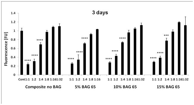
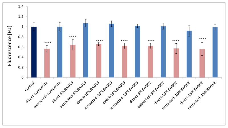
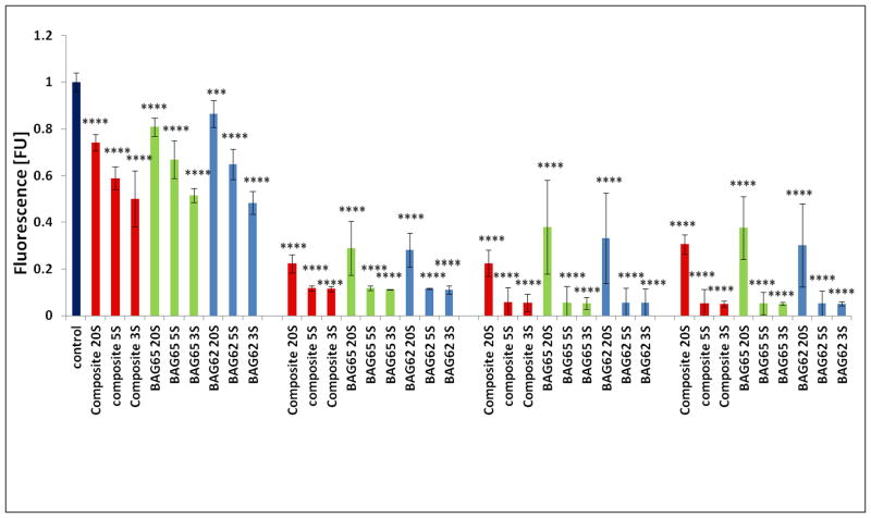
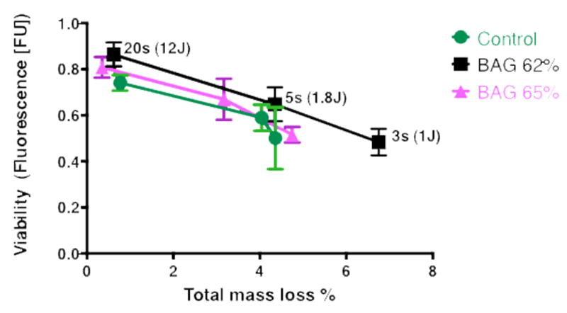
Dental composites (50:50 Bis-GMA/TEGDMA resin: 72.5wt% filler, 67.5%Sr-glass and 5% OX50) containing different concentrations (0, 5, 10 and 15wt%) of two sol-gel bioactive glasses, BAG65 (65mole% SiO2, 31mole% CaO, 4mole% P2O5) and BAG61 (3mole% F added) were evaluated for cytotoxicity using Alamar Blue assay. First, composite extracts were obtained from 7 day incubations of composites in cell culture medium at 37°C. Undifferentiated pulp cells (OD-21) were exposed to dilutions of the original extracts for 3, 5, and 7 days. Then freshly cured composite disks were incubated with OD-21 cells (n=5) for 2 days. Subsequently, fresh composite disks were incubated in culture medium at 37°C for 7 days, and then the extracted disks were incubated with OD-21 cells for 2 days. Finally, fresh composites disks were light cured for 3, 5, and 20s and incubated with OD-21 cells (n=5) for 1, 3, 5, and 7 days. To verify that the three different curing modes produced different levels of degree of conversion (DC), the DC of each composite was determined by FTIR. Groups (n=5) were compared with ANOVA/Tukey's (α≤0.05).

RESULTS

Extracts from all composites significantly reduced cell viability until a dilution of 1:8 or lower, where the extract became equal to the control. All freshly-cured composites showed significantly reduced cell viability at two days. However, no reduction in cell viability was observed for any composite that had been previously soaked in media before exposure to the cells. Composites with reduced DC (3s vs. 20s cure), as verified by FTIR, showed significantly reduced cell viability.

SIGNIFICANCE

The results show that the composites, independent of composition, had equivalent potency in terms of reducing the viability of the cells in culture. Soaking the composites for 7 days before exposing them to the cells suggested that the "toxic" components had been extracted and the materials were no longer cytotoxic. The results demonstrate that the cytotoxicity of composites with and without BAG must predominantly be attributed to the release of residual monomers, and not to the presence of the BAG.

摘要

目的

测定含生物活性玻璃填料的牙科复合材料的体外细胞毒性。

方法

使用alamar蓝测定法评估含有两种溶胶 - 凝胶生物活性玻璃BAG65(65摩尔%二氧化硅,31摩尔%氧化钙,4摩尔%五氧化二磷)和BAG61(添加3摩尔%氟)不同浓度(0、5、10和15重量%)的牙科复合材料(50:50双酚A - 双甲基丙烯酸缩水甘油酯/三乙二醇二甲基丙烯酸酯树脂:72.5重量%填料,67.5%锶玻璃和5%OX50)的细胞毒性。首先,通过在37°C的细胞培养基中对复合材料进行7天孵育获得复合提取物。将未分化的牙髓细胞(OD - 21)暴露于原始提取物的稀释液中3、5和7天。然后将新固化的复合圆盘与OD - 21细胞(n = 5)孵育2天。随后,将新鲜的复合圆盘在37°C的培养基中孵育7天,然后将提取的圆盘与OD - 21细胞孵育2天。最后,将新鲜的复合圆盘光固化3、5和20秒,并与OD - 21细胞(n = 5)孵育1、3、5和7天。为了验证三种不同的固化模式产生不同程度的转化率(DC),通过傅里叶变换红外光谱(FTIR)测定每种复合材料的DC。使用方差分析/图基检验(α≤0.05)对组(n = 5)进行比较。

结果

所有复合材料的提取物在稀释至1:8或更低之前均显著降低细胞活力,此时提取物与对照相当。所有新固化的复合材料在两天时均显示细胞活力显著降低。然而,对于任何在暴露于细胞之前已在培养基中浸泡过的复合材料,未观察到细胞活力降低。经FTIR验证,转化率降低的复合材料(3秒与20秒固化)显示细胞活力显著降低。

意义

结果表明,复合材料无论其组成如何,在降低培养细胞活力方面具有同等效力。在将复合材料暴露于细胞之前将其浸泡7天表明“有毒”成分已被提取,材料不再具有细胞毒性。结果表明,含或不含生物活性玻璃的复合材料的细胞毒性主要归因于残留单体的释放,而非生物活性玻璃的存在。

https://cdn.ncbi.nlm.nih.gov/pmc/blobs/8399/4448918/17740ed1fc76/nihms653696f5.jpg
https://cdn.ncbi.nlm.nih.gov/pmc/blobs/8399/4448918/102e08fa9178/nihms653696f1a.jpg
https://cdn.ncbi.nlm.nih.gov/pmc/blobs/8399/4448918/dc7a09487e9d/nihms653696f2.jpg
https://cdn.ncbi.nlm.nih.gov/pmc/blobs/8399/4448918/2bdab4066d3e/nihms653696f3.jpg
https://cdn.ncbi.nlm.nih.gov/pmc/blobs/8399/4448918/20adae75e260/nihms653696f4.jpg
https://cdn.ncbi.nlm.nih.gov/pmc/blobs/8399/4448918/17740ed1fc76/nihms653696f5.jpg