Dhupia Neha, Rathour Rahul K, Narayanan Rishikesh
Cellular Neurophysiology Laboratory, Indian Institute of Science Bangalore, India ; Centre for Converging Technologies, University of Rajasthan Jaipur, India.
Cellular Neurophysiology Laboratory, Indian Institute of Science Bangalore, India.
Front Cell Neurosci. 2015 Jan 12;8:456. doi: 10.3389/fncel.2014.00456. eCollection 2014.
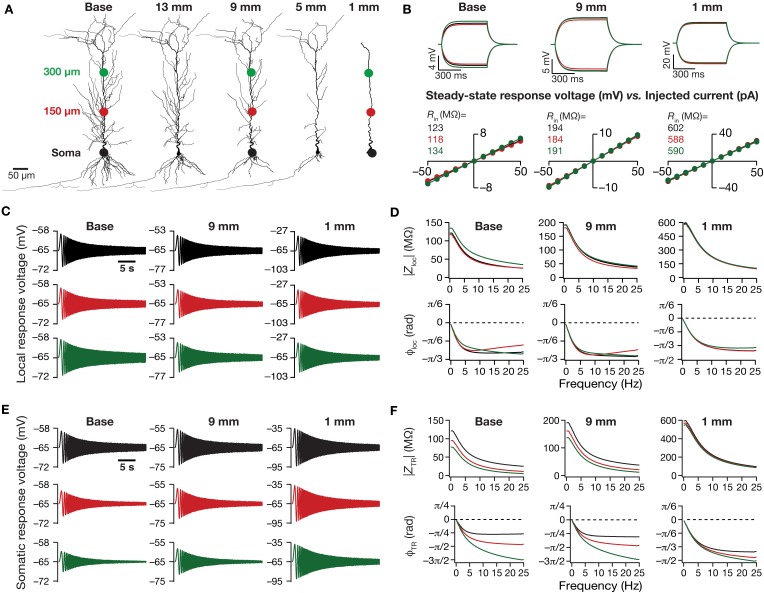
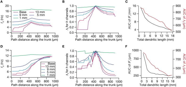
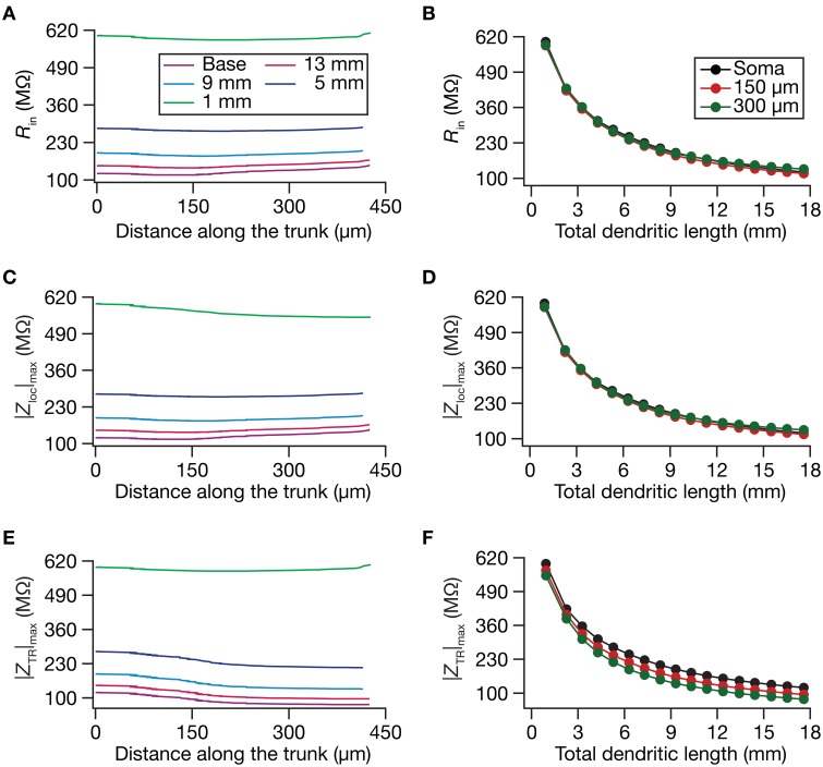
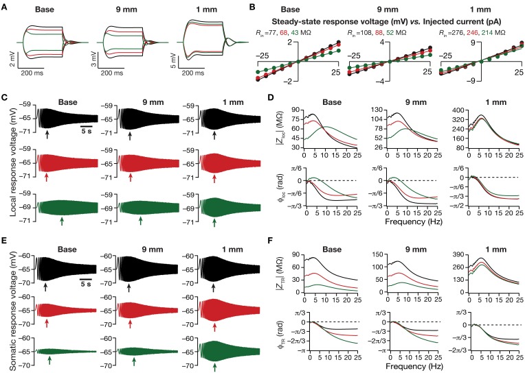
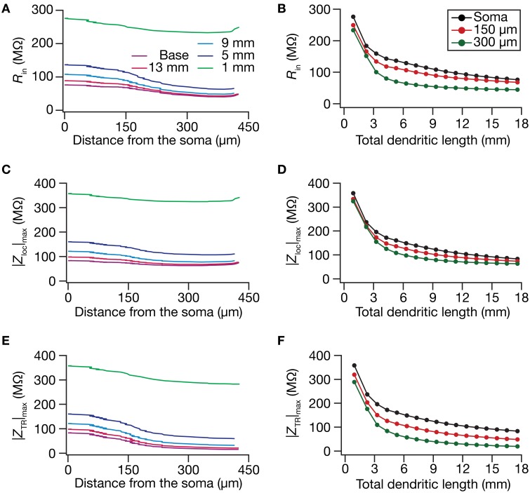
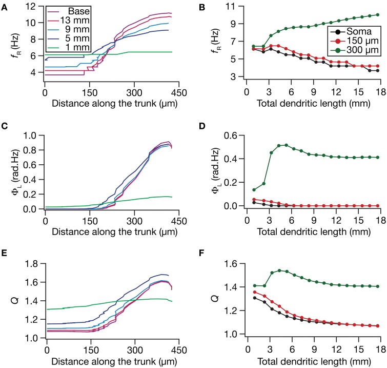
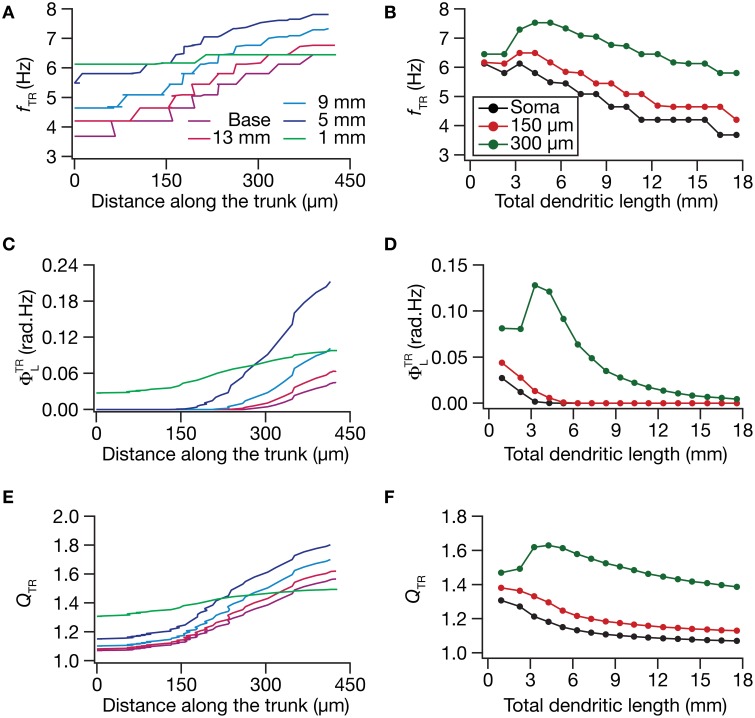
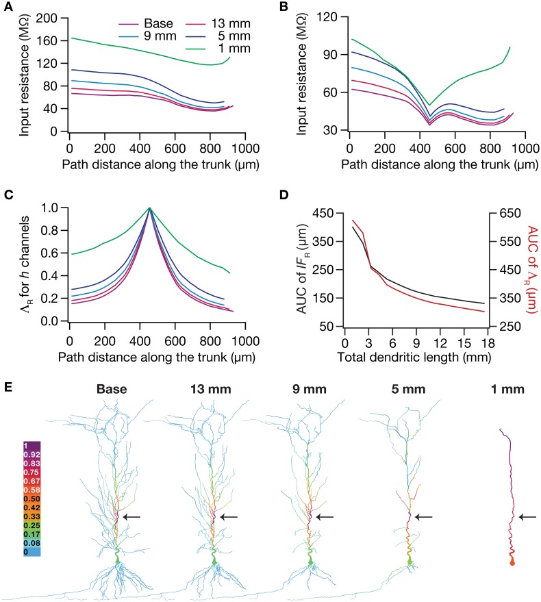
A gradient in the density of hyperpolarization-activated cyclic-nucleotide gated (HCN) channels is necessary for the emergence of several functional maps within hippocampal pyramidal neurons. Here, we systematically analyzed the impact of dendritic atrophy on nine such functional maps, related to input resistance and local/transfer impedance properties, using conductance-based models of hippocampal pyramidal neurons. We introduced progressive dendritic atrophy in a CA1 pyramidal neuron reconstruction through a pruning algorithm, measured all functional maps in each pruned reconstruction, and arrived at functional forms for the dependence of underlying measurements on dendritic length. We found that, across frequencies, atrophied neurons responded with higher efficiency to incoming inputs, and the transfer of signals across the dendritic tree was more effective in an atrophied reconstruction. Importantly, despite the presence of identical HCN-channel density gradients, spatial gradients in input resistance, local/transfer resonance frequencies and impedance profiles were significantly constricted in reconstructions with dendritic atrophy, where these physiological measurements across dendritic locations converged to similar values. These results revealed that, in atrophied dendritic structures, the presence of an ion channel density gradient alone was insufficient to sustain homologous functional maps along the same neuronal topograph. We assessed the biophysical basis for these conclusions and found that this atrophy-induced constriction of functional maps was mediated by an enhanced spatial spread of the influence of an HCN-channel cluster in atrophied trees. These results demonstrated that the influence fields of ion channel conductances need to be localized for channel gradients to express themselves as homologous functional maps, suggesting that ion channel gradients are necessary but not sufficient for the emergence of functional maps within single neurons.
超极化激活的环核苷酸门控(HCN)通道密度梯度对于海马锥体细胞内多个功能图谱的出现是必要的。在此,我们使用基于电导的海马锥体细胞模型,系统分析了树突萎缩对九个此类功能图谱的影响,这些图谱与输入电阻以及局部/传递阻抗特性有关。我们通过修剪算法在CA1锥体细胞重建中引入渐进性树突萎缩,测量每个修剪后重建中的所有功能图谱,并得出基础测量值对树突长度依赖性的函数形式。我们发现,在各个频率下,萎缩的神经元对传入输入的响应效率更高,并且在萎缩的重建中,信号在树突树上的传递更有效。重要的是,尽管存在相同的HCN通道密度梯度,但在树突萎缩的重建中,输入电阻、局部/传递共振频率和阻抗分布的空间梯度显著收缩,在这些重建中,跨树突位置的这些生理测量值收敛到相似的值。这些结果表明,在萎缩的树突结构中,仅离子通道密度梯度的存在不足以维持沿相同神经元拓扑图的同源功能图谱。我们评估了这些结论的生物物理基础,发现这种萎缩诱导的功能图谱收缩是由萎缩树中HCN通道簇影响的空间扩散增强介导的。这些结果表明,离子通道电导的影响场需要定位,以便通道梯度表现为同源功能图谱,这表明离子通道梯度对于单个神经元内功能图谱的出现是必要但不充分的。